
Fig. S12
- ID
- ZDB-IMAGE-110214-33
- Publication
- Tittle et al., 2011 - Uhrf1 and Dnmt1 are required for development and maintenance of the zebrafish lens
- All Figures
- Figures for Tittle et al., 2011
|
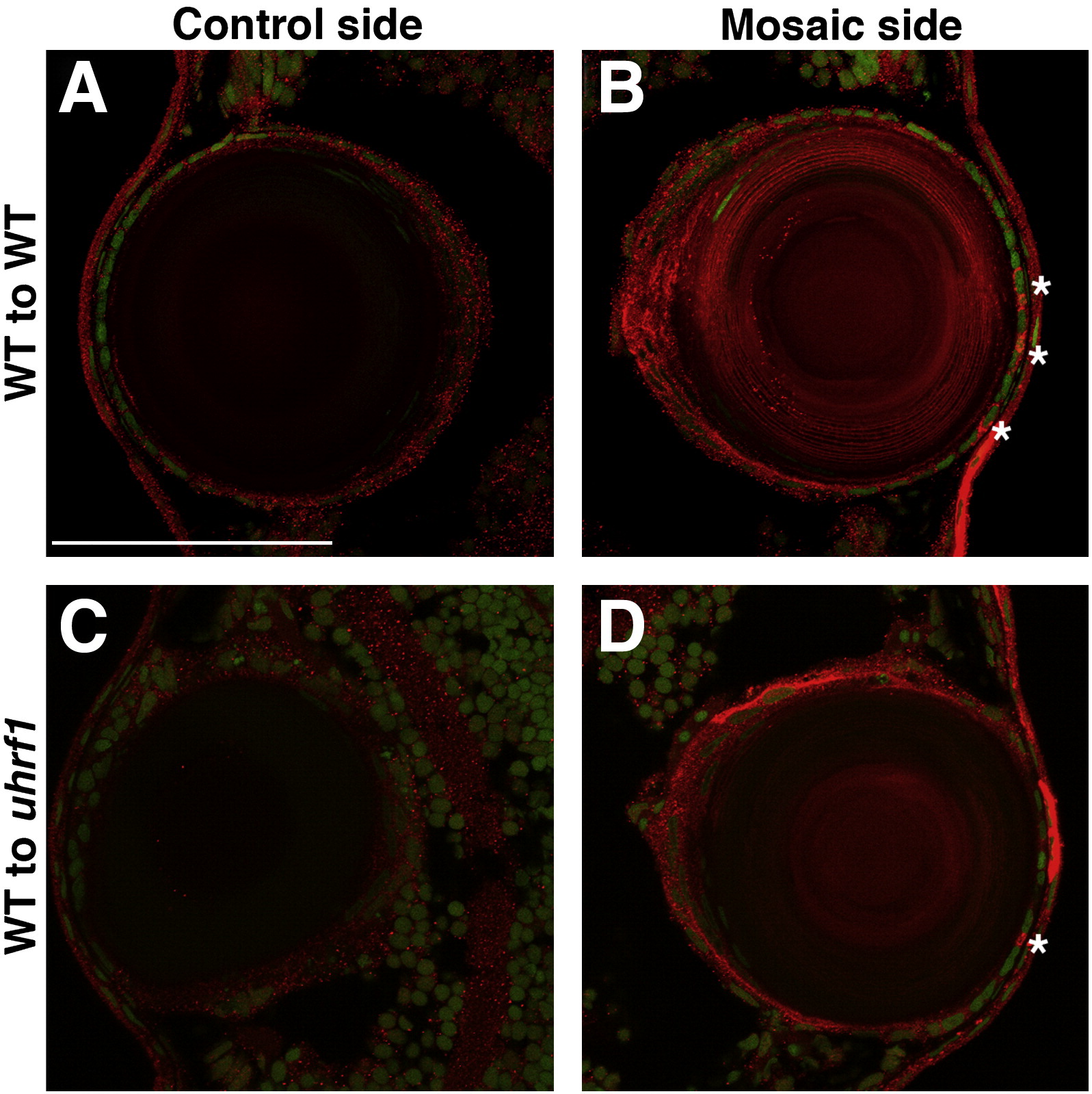
Fig. S12 Wild-type donor cells do not outcompete and repopulate the lens epithelium of a uhrf1 host. Shield-stage transplants were performed in which donor cells were derived from transgenic zebrafish expressing beta actin2:mCherry. uhrf1 mutants or phenotypically wild-type siblings were the transplant hosts. The resulting mosaic embryos were cryosectioned, stained with an anti-dsRed antibody, and imaged by confocal microscopy to visualize beta actin2:mCherry-expressing donor-derived cells within the host lens. (A,B) control (wild-type donor → wild-type host) or (C,D) experimental (wild-type donor → uhrf1 host) mosaic lenses at 4 dpf. In both cases, the lens epithelium contained only a few donor-derived cells. For each mosaic embryo, both the non-transplanted (fully host-derived) lens (A,C), and the mosaic lens (B,D) are shown. Scale bars are 70 μm.
Reprinted from Developmental Biology, 350(1), Tittle, R.K., Sze, R., Ng, A., Nuckels, R.J., Swartz, M.E., Anderson, R.M., Bosch, J., Stainier, D.Y., Eberhart, J.K., and Gross, J.M., Uhrf1 and Dnmt1 are required for development and maintenance of the zebrafish lens, 50-63, Copyright (2011) with permission from Elsevier. Full text @ Dev. Biol.