
|
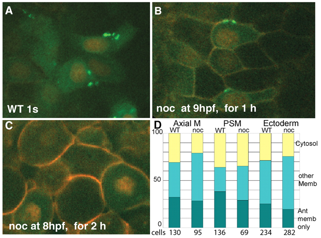
Fig. 5 Intact microtubules are not required for Pk maintenance at anterior cell membrane, but are need for establishment of anterior localization. In all panels, embryos are one-somite stage and anterior is upwards; cells are presumptive mesoderm (at least one cell layer away from the enveloping layer cells or clearly below ectoderm/mesoderm boundary). Cells are labeled with Histone2B-RFP, Membrane-RFP and Pk-GFP. (A) Wild-type embryo grown without nocodazole, (B) grown in 10 μg/ml nocodazole for 1 hour, starting at late gastrulation or (C) grown in 10 μg/ml nocodazole for 2 hours, starting at midgastrulation. (D) The location of Pk-GFP labeling was noted for axial mesoderm, presomitic mesoderm (PSM) and ectoderm in wild-type and 1 hour nocodazole-treated embryos (n=7, each condition). Cellular localization is largely stable to 1 hour treatment.