
Image
|
Figure Caption
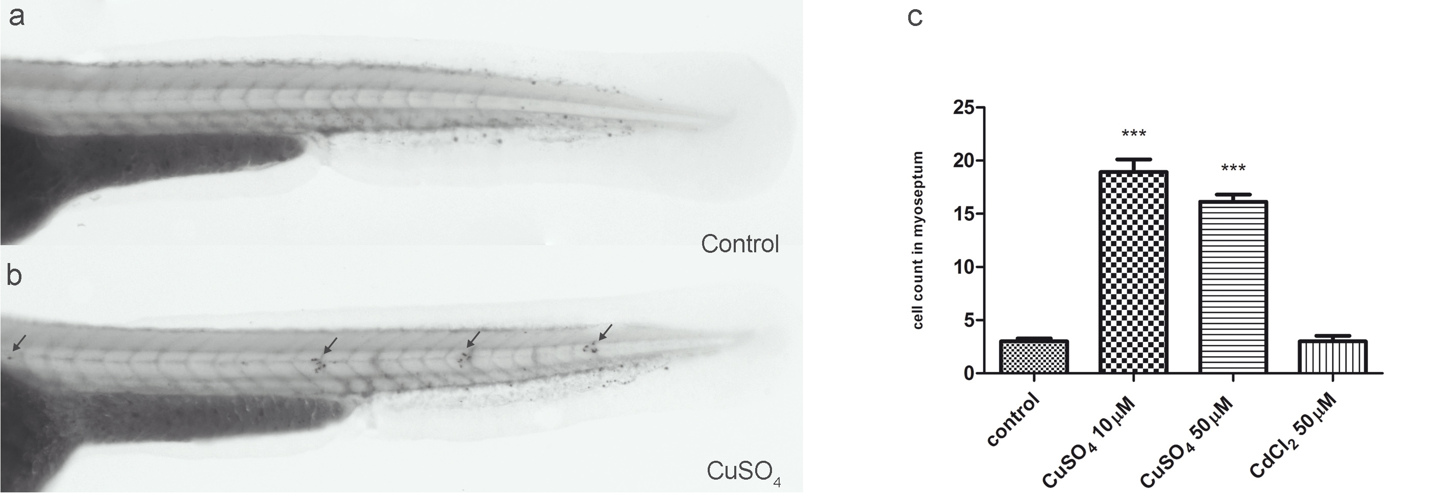
Fig. S4 Chemically induced inflammation assay (ChIn) using Sudan Black. (a and b) Bright-field images of untreated (a) and 10 μM CuSO4-treated (b) 56-hpf casper larvae stained with Sudan Black to reveal leukocytes. Note the congregation of labeled cells at the posterior lateral line neuromasts (arrows). (c) Quantification of leukocyte migration (detected by Sudan Black staining) to the lateral line in untreated and metal-exposed larvae. The result is equivalent to that obtained with BACmpx::GFP or lysC::DsRED2 transgenic larvae.
Acknowledgments
This image is the copyrighted work of the attributed author or publisher, and
ZFIN has permission only to display this image to its users.
Additional permissions should be obtained from the applicable author or publisher of the image.
Open Access.
Full text @ BMC Biol.