
Fig. 1
- ID
- ZDB-IMAGE-110121-14
- Genes
- Publication
- Zhong et al., 2011 - SSDP cofactors regulate neural patterning and differentiation of specific axonal projections
- All Figures
- Figures for Zhong et al., 2011
|
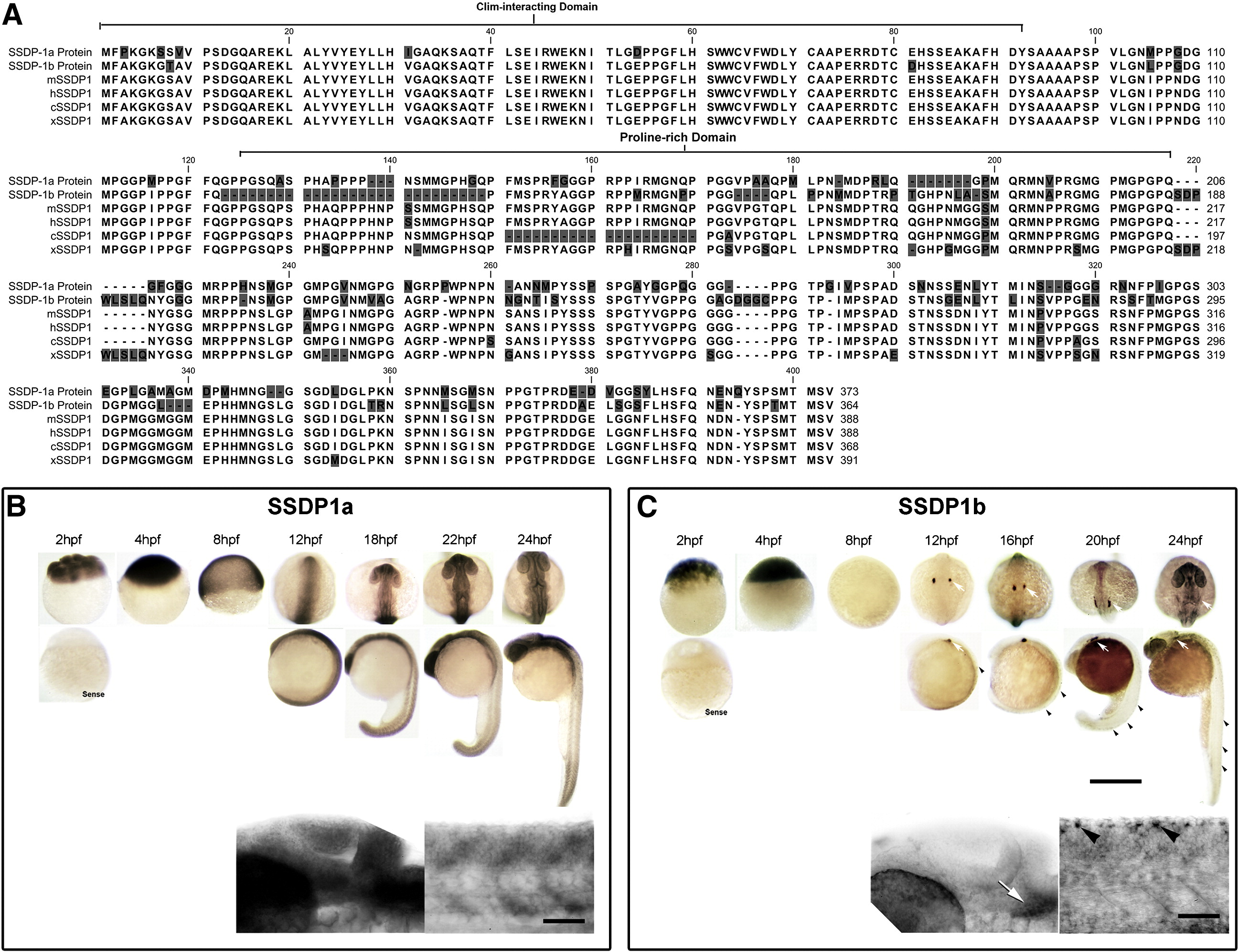
Fig. 1 SSDP1a and SSDP1b protein sequences are conserved and mRNAs are expressed during embryonic development in zebrafish. A: The CLIM-interacting domain is most strongly conserved between zebrafish SSDP1s and the mouse (mSSDP1), human (hSSDP1), chick (cSSDP1) and Xenopus (xSSDP1) protein. B, C: Whole-mount in situ hybridization between 2 and 24 hpf indicates that SSDP1a is ubiquitously expressed with stronger expression in the head, whereas SSDP1b mRNA is most strongly expressed in trigeminal (white arrows) and Rohon–Beard (black arrowheads) neurons. Higher magnification pictures in lower row show lateral views of the head (left) and trunk (right). Upper scale bar in C = 500 μm for low magnification pictures; lower scale bar = 50 μm for high magnification pictures.
Reprinted from Developmental Biology, 349(2), Zhong, Z., Ma, H., Taniguchi-Ishigaki, N., Nagarajan, L., Becker, C.G., Bach, I., and Becker, T., SSDP cofactors regulate neural patterning and differentiation of specific axonal projections, 213-224, Copyright (2011) with permission from Elsevier. Full text @ Dev. Biol.