
Fig. 6
- ID
- ZDB-IMAGE-110112-14
- Publication
- Schwend et al., 2010 - Visualization of gli activity in craniofacial tissues of hedgehog-pathway reporter transgenic zebrafish
- All Figures
- Figures for Schwend et al., 2010
|
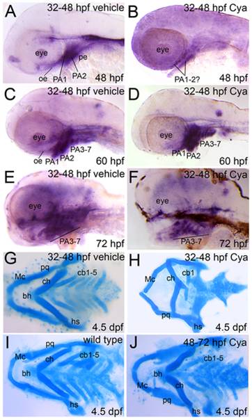
Fig. 6 Reduced Gli activity in craniofacial tissues following late pharyngula stage Hh-pathway inhibition.
Lateral (A–F) or dorsal (G–J) views, anterior to the left, of Tg(Gli-d:mCherry) fish treated with vehicle (A,C,E,G), Cya (B,D,F,H,J) or not treated (I) and stained with mCherry riboprobe (A–F) or Alcian blue solution (G–J). (A,B) Reporter gene expression was significantly reduced in the brain and facial epithelium at 48 hpf following late pharyngula stage Cya treatment. A slight mCherry signal was detected in craniofacial tissue that likely corresponds to PA1-2 mesenchyme at 48 hpf following Cya treatment. (C–F) Reporter gene expression was fully recovered in Cya treated embryos by 60–72 hpf and is expressed in oe, pe, brain and PA mesenchyme. (M–O) Alcian blue staining to reveal cartilage deficits in Gli reporter transgenic fish following Cya treatments at 32–48 hpf (H) or 48–72 hpf (J). These deficits are consistent with our previously published findings and indicate that Cya reliably inhibited cb chondrofication upon Hh-inhibition during the late pharyngula stage (H), but did not negatively impact cartilage development when administered at later stages (J).