
Fig. S3
- ID
- ZDB-IMAGE-101223-29
- Publication
- Lee et al., 2010 - The habenula prevents helpless behavior in larval zebrafish
- All Figures
- Figures for Lee et al., 2010
|
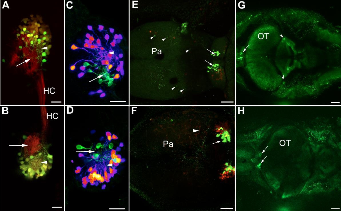
Fig. S3 Related to Figure 4. Kaede and TeTXlc-CFP Expression in GAL4s1019t Fish
(A and B) Dorsal view of the right (A) and left (B) habenula of a KR11/GAL4s1019t/UAS:Kaede larva. Habenula neurons expressing Kaede (green) extend projections into dorsal habenula neuropils that are innervated by KillerRed expressing axons (arrowheads). The most medial neuropil (arrow) contains very few Kaede-expressing fibers.
(C and D) Dorsal view of the right (C) and left (D) habenula of GAL4s1019t/UAS:Kaede/UAS:TeTXlc-CFP fish, examined after conditioning. TeTXlc-CFP neurons (green) extend processes into a more medial location than Kaede-expressing neurons (purple-red-orange).
(E-H) Low magnification of GAL4s1019t/UAS:Kaede/UAS:TeTXlc-CFP fish, examined after conditioning.
(E and F) TeTXlc-CFP (green; arrowheads) and Kaede (red) are visible in several forebrain neurons.
(F) Kaede is expressed in pericytes that associated with vasculature (arrowhead) in this fish. TeTXlc-CFP was detected in pericytes in one fish.
(G) TeTXlc-CFP is visible in neurons located in the eminentia granulosum of the cerebellum (arrowheads) of this fish. Similar expression was seen in 3 other fish.
(H) A fish with no TeTXlc-CFP in the cerebellum. Speckles outside the CNS are due to scatter from the skin. The arrows indicate expression in the medial region of the dorsal habenula.
Pa: pallium; OT: optic tectum; HC: habenular commissure. Anterior is to the left for all images. Bar = 20 μm for panels A-D, 50 μm for panels E, F and 100 μm for panels G, H. All images are projections of z-stacks.