
IMAGE
Fig. 3
- ID
- ZDB-IMAGE-101223-15
- Genes
- Antibodies
- Publication
- Santoriello et al., 2010 - Kita Driven Expression of Oncogenic HRAS Leads to Early Onset and Highly Penetrant Melanoma in Zebrafish
- All Figures
- Figures for Santoriello et al., 2010
Image
|
Figure Caption
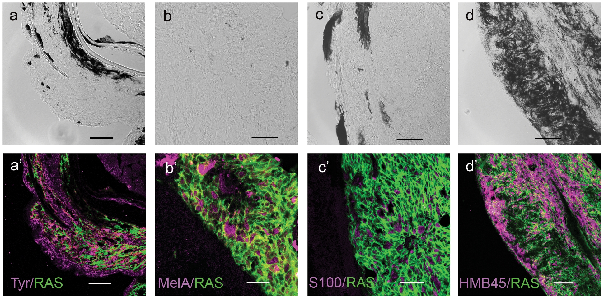
Fig. 3 Immunostaining of zebrafish melanoma cryostat sections.
In the lower panels (a′–d′) immunostainings (magenta) for the markers indicated in the lower right corners of each micrograph. All cells express GFP-RAS (green at the plasma membrane). Upper panels (a–d) show the DIC image of each sample, which consisted mostly of unpigmented tumors. Calibration bars = 50 μm.
Figure Data
Acknowledgments
This image is the copyrighted work of the attributed author or publisher, and
ZFIN has permission only to display this image to its users.
Additional permissions should be obtained from the applicable author or publisher of the image.
Full text @ PLoS One