
|
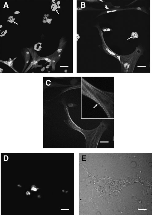
Fig. 4 Expression of transgenic markers in seZEB cultures. Single-embryo cell cultures were established using embryos from unc45b-GFP or fli1-GFP transgenic zebrafish, which fluoresce in muscle tissue and endothelial tissue, respectively. (A, B)) Expression of unc45b-GFP was limited to bundles of differentiating myocytes, while isolated cells (indicated by DAPI staining) at the edges of the culture (A) or between the myocyte bundles (B) displayed no detectable GFP (arrows). (C) Actin staining with Alexa 568 phalloidin revealed that GFP-positive cells also displayed actin banding patterns characteristic of myocytes (arrow). Inset shows higher magnification. (D) Expression of fli1-GFP was not detected in any culture, although differentiating cells still appeared flattened and elongated like wild-type (WT) cultures under phase-contrast microscopy (E). All cultures examined contained visibly differentiating myocytes which were positive for unc45b-GFP expression (n = 40 single-embryo cultures). Scale bars in all panels = 0.1mm.