
|
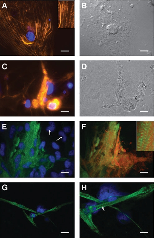
Fig. 3 Molecular characterization of cells derived from seZEB cultures. Single-embryo cultures were stained for several markers of myocyte differentiation. Actin expression in seZEB cultures is shown by Alexa 546-phalloidin staining (A, C) (orange), whereas nuclei are indicated by DAPI staining (blue). Single-nucleated myocytes (A, inset) and multinucleated myocyte bundles (C) both demonstrate distinct banded patterns of actin expression. (B, D) Phase-contrast images of the stained cultures from A and C, demonstrating a single cell body (A) versus a bundle of elongated cells (C). (E) Muscle myosin was detected using F-59 anti-myosin heavy chain antibody and Alexa 488 anti-mouse secondary antibody (green), with DAPI counterstain (blue). Myosin-negative cells are indicated with arrows. (F) Myosin and actin costained cultures, showing alternating patterns of myosin and actin sarcomere bands characteristic of differentiating muscle. Higher magnification is shown in the inset. Comparison with cultures of differentiated tail myocytes from 3-day-old zebrafish embryos demonstrated a similar phenotype (G) and pattern of muscle myosin expression (H) in characteristic M-bands (arrow). Scale bars = 0.05mm (A–H) and 0.1mm (G).