
Fig. 6
- ID
- ZDB-IMAGE-101208-29
- Publication
- Schneider et al., 2010 - Zebrafish Nkd1 promotes Dvl degradation and is required for left-right patterning
- All Figures
- Figures for Schneider et al., 2010
|
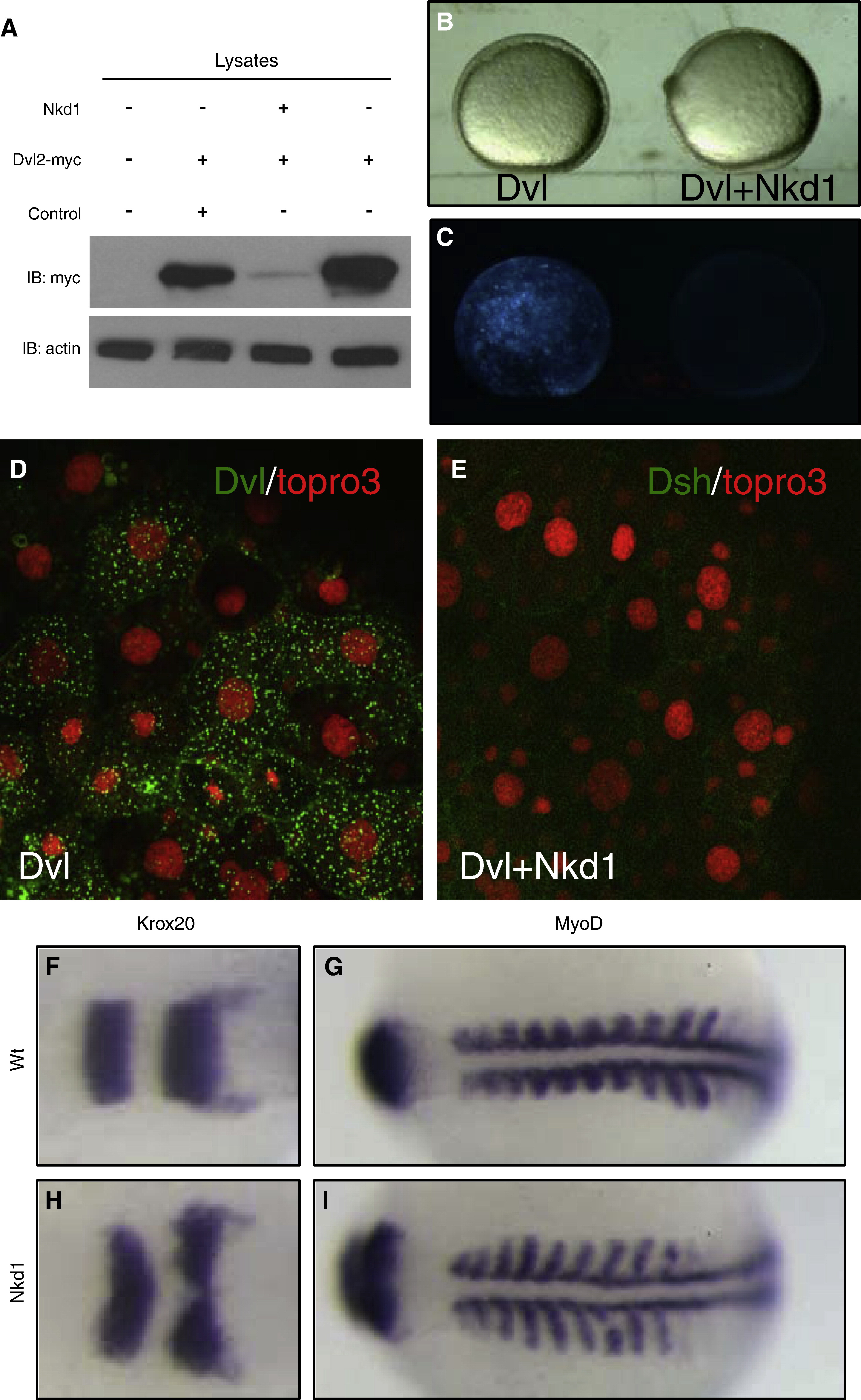
Fig. 6 Nkd1 promotes Dvl degradation and impacts CE movements. (A) Westerm blot of zebrafish embryos injected with myc-tagged Dvl2 only, or coinjected with either nkd1 RNA or egfp RNA. The first lane is uninjected embryos, anti-β-actin was used as loading control. Embryos were frozen at 80% epiboly and equal amounts of cell lysates were used for Western blot analysis. (B) Bright field and (C) fluorescence images of embryos at 90% epiboly, injected with Dvl-GFP only (left) or coinjected with nkd1 RNA (right). (D) Dvl-GFP (green) and Topro3 nuclear staining (red) denotes Dvl localization in wt and (E) Dvl-GFP coinjected with nkd1 RNA. (F) krox20 and (G) myoD markers in uninjected and (H) krox20 and (I) myoD markers nkd1 RNA-injected zebrafish embryos. 43% of nkd1 injected embryos show CE defects, n = 143. Dorsal view, anterior to the left.
Reprinted from Developmental Biology, 348(1), Schneider, I., Schneider, P.N., Derry, S.W., Lin, S., Barton, L.J., Westfall, T., and Slusarski, D.C., Zebrafish Nkd1 promotes Dvl degradation and is required for left-right patterning, 22-33, Copyright (2010) with permission from Elsevier. Full text @ Dev. Biol.