
IMAGE
Fig. S2
- ID
- ZDB-IMAGE-101207-15
- Publication
- Martin et al., 2010 - Wallerian degeneration of zebrafish trigeminal axons in the skin is required for regeneration and developmental pruning
- All Figures
- Figures for Martin et al., 2010
Image
|
Figure Caption
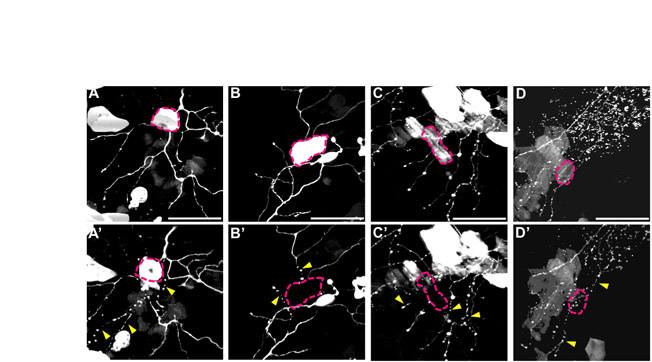
Fig. S2 Keratinocyte damage causes immediate axon fragmentation following axotomy. Two-photon image stack projections of four different trigeminal axons (A-D) and neighboring keratinocytes expressing GFP. (A-D) Skin cells (marked by pink dotted lines) that overlap with axons before axotomy and ablation. Two-photon laser ablation of skin cells was performed concurrently with axotomy of the adjacent axon. (A′-D′) Immediately after ablation/axotomy. Pink dotted lines indicate damaged/dead keratinocytes, and yellow arrowheads indicate axons that instantly fragmented (n=5). Scale bars: 50 μm.
Acknowledgments
This image is the copyrighted work of the attributed author or publisher, and
ZFIN has permission only to display this image to its users.
Additional permissions should be obtained from the applicable author or publisher of the image.
Full text @ Development