
Image
|
Figure Caption
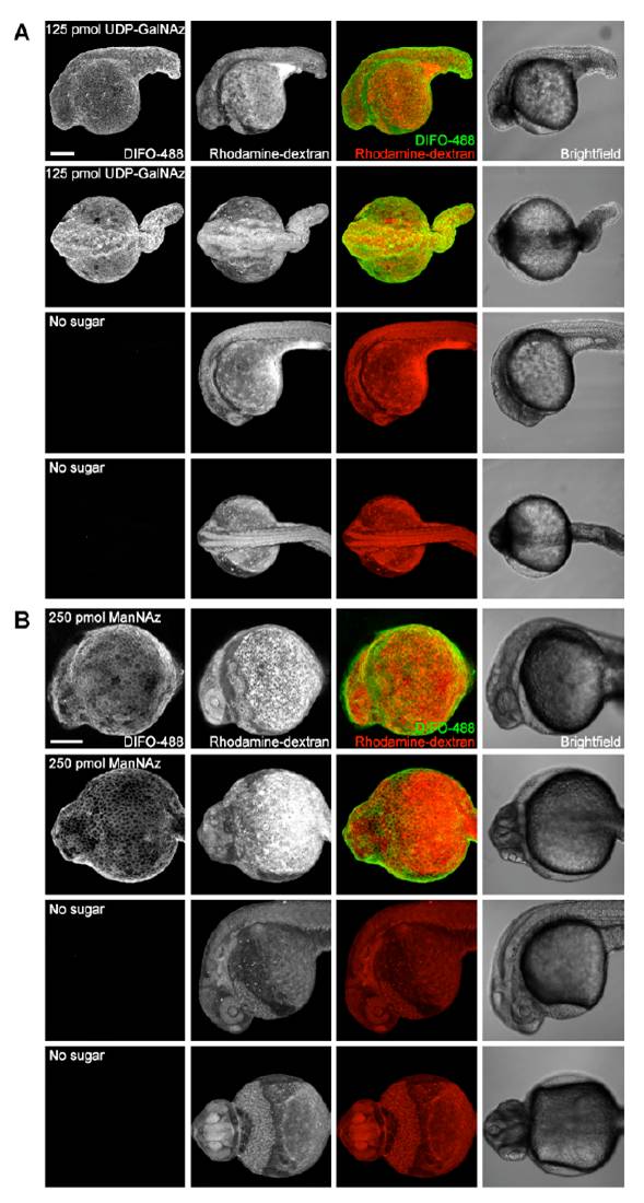
Fig. S3 Microinjection of higher doses of azidosugars. Zebrafish embryos were microinjected with 125 pmol of UDP-GalNAz (A, top), 125 pmol of UDP-GalNAc (A, bottom), 250 pmol of ManNAz (B, top), or no sugar (B, bottom), along with the tracer dye rhodaminedextran. The embryos were allowed to develop to 24 hpf, reacted with DIFO-488 (100 μM, 1 h), and imaged by confocal microscopy. Shown are maximum intensity z-projection fluorescence images and corresponding brightfield images. Green, DIFO-488; red, rhodamine-dextran. Scale bar: 200 μm.
Acknowledgments
This image is the copyrighted work of the attributed author or publisher, and
ZFIN has permission only to display this image to its users.
Additional permissions should be obtained from the applicable author or publisher of the image.
Full text @ Proc. Natl. Acad. Sci. USA