
Image
|
Figure Caption
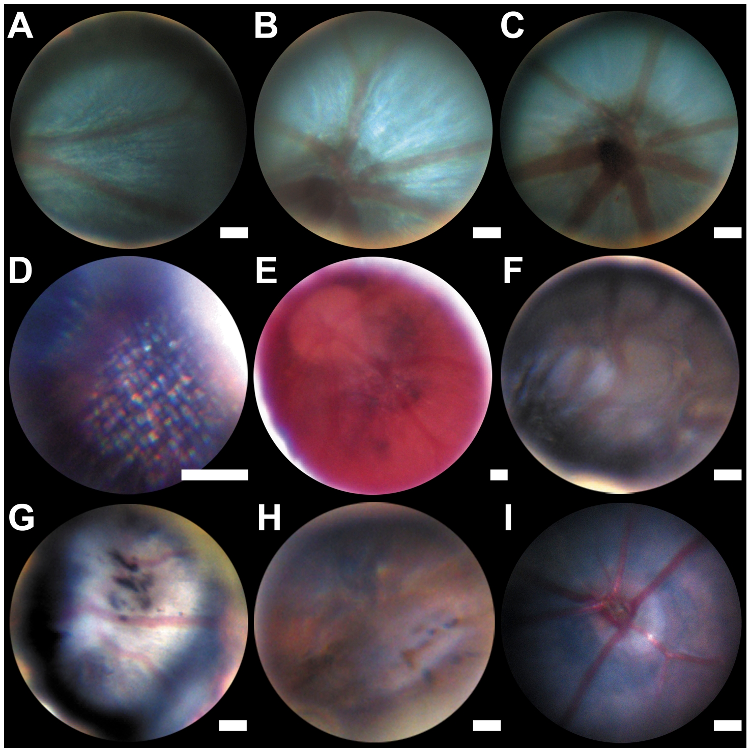
Fig. 3 Zebrafish and mouse fundus images.
(A–D) wild type zebrafish fundus at the level of the mid-peripheral retina (A), optic disc (B and C) and photoreceptor level, showing the presumed photoreceptor mosaic (D). Hypopigmented fundus of the zebrafish albino strain (E). Tortuous arteries and a darkened retina (F), black spots are visible in the retina (G and H). Fundus Image of a wild type mouse (I). Estimated scale bar: 50 μm.
Figure Data
Acknowledgments
This image is the copyrighted work of the attributed author or publisher, and
ZFIN has permission only to display this image to its users.
Additional permissions should be obtained from the applicable author or publisher of the image.
Full text @ PLoS One