
Fig. 1
- ID
- ZDB-IMAGE-101123-51
- Publication
- Chung et al., 2010 - Suppression of Alk8-mediated Bmp signaling cell-autonomously induces pancreatic β-cells in zebrafish
- All Figures
- Figures for Chung et al., 2010
|
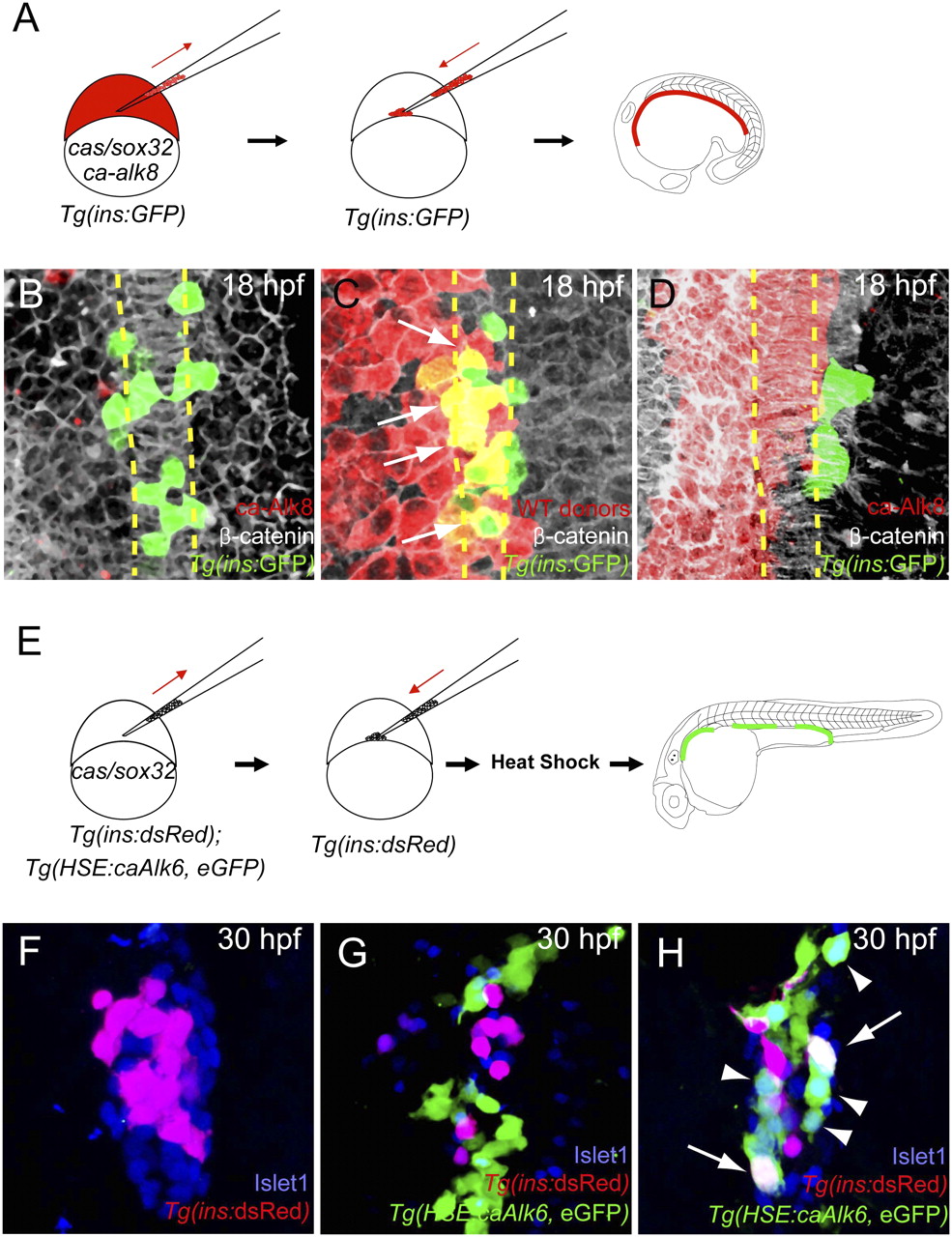
Fig. 1
Activation of Bmp signaling cell-autonomously blocks the induction of dorsal bud-derived endocrine cells. (A) Schematic diagram of the cell transplantation protocol. Tg(ins:GFP) donors were injected with cas/sox32 and ca-alk8 RNA, along with rhodamine dextran, and the cells were transplanted into Tg(ins:GFP) hosts. (B–D) Ventral confocal images of Tg(ins:GFP) (green), β-catenin (white), and rhodamine dextran (red) at 18 hpf (the notochord is outlined by yellow dashed lines). (B and C) Tg(ins:GFP)-expressing cells are normally located close to the notochord in wild type (B) and hosts containing control donor cells (C). (D) Tg(ins:GFP) expression was absent in ca-Alk8-expressing endodermal cells. (E) Schematic diagram of the cell transplantation protocol. cas/sox32 overexpressing Tg(ins:dsRed); Tg(HSE:caAlk6, eGFP) donor cells were transplanted into Tg(ins:dsRed) hosts, and subsequently heat-shocked at various time points. (F–H) Confocal projection images of hosts with Tg(HSE:caAlk6, eGFP)-expressing cells (green) at 30 hpf after staining for dsRed (red) and Islet1 (blue), comparing control (F) and hosts [heat shock was applied at 9 (G) or 11 hpf (H)]. (G) When ca-alk6 expression was induced at 9 hpf, the number of Tg(ins:dsRed) and Islet1-expressing cells was reduced (compare F and G), and the Tg(HSE:caAlk6, eGFP)-expressing cells failed to express endocrine markers. (H) When ca-alk6 expression was induced at 11 hpf, several Tg(HSE:caAlk6, eGFP)-expressing cells expressed Islet1 (arrowheads) and some of them also coexpressed Tg(ins:dsRed) (arrows).