
Fig. S4
- ID
- ZDB-IMAGE-101119-6
- Genes
- Publication
- Friedman et al., 2009 - MicroRNAs are essential for development and function of inner ear hair cells in vertebrates
- All Figures
- Figures for Friedman et al., 2009
|
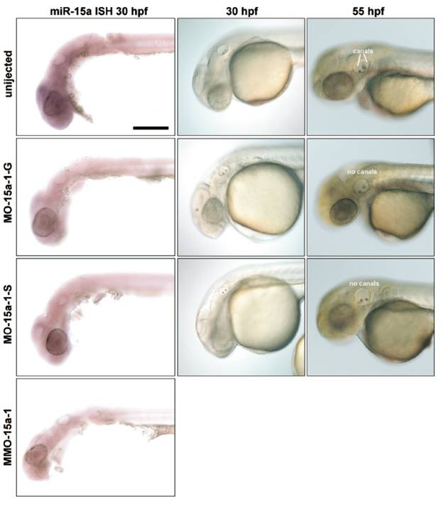
Fig. S4 Morpholino knockdown of miR-15a-1 using different MOs. (Left) Thirty-hpf embryos probed with miR-15a locked nucleic acid (LNA) probe to detect mature miRNAs. (Scale bar, 200 μm.) (Middle) Nomarski images of embryos at 30 hpf show presence of 2 otoliths in the inner ear; MMO-15a-1 morphants are indistinguishable from uninjected embryos (data not shown). (Right) Nomarski images of embryos at 55 hpf showing that miR-15a-1 morphants fail to show proper initiation of semicircular canal formation (image of MMO-15a-1 morphant ear at this stage is shown in Fig. 4B and Fig. S6B). Many embryos treated with MO-15a-1-S look normal at this stage; the embryo shown is representative of fewer than 10% that show the canal phenotype.