IMAGE

Fig. S1

ID
ZDB-IMAGE-101118-46
Source
Figures for Hesselson et al., 2009
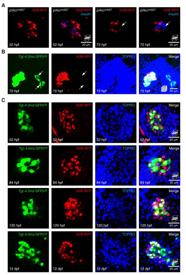
Image
Figure Caption

Fig. S1 H2B-RFP is retained in dorsal bud derived 7beta;-cells during embryonic and larval development. All embryos were injected with H2B-RFP mRNA at the one cell stage. (A) Confocal sections of prkci mutants, at 52 and 72 hpf, stained with insulin. (B) Confocal projection of an islet stained with TOPRO. (C) Confocal sections of islets that express Tg(-4.0ins:GFP)zf5 at different developmental time-points stained with TOPRO. (A) All insulin-expressing cells retain the H2B-RFP label at 52 hpf in prkcim567 mutants. By 72 hpf, label diluted VBCs appear (arrow). (B) Tg(-4.0ins:GFP)zf5-positive cells (arrows) that differentiate outside the principal islet do not exhibit the H2B-RFP label. (C) H2B-RFP label-retaining endodermal cells can be clearly distinguished from the surrounding label-diluted cells by 52 hpf. The H2B-RFP label remains easily detectable at 12 dpf.

Acknowledgments
This image is the copyrighted work of the attributed author or publisher, and ZFIN has permission only to display this image to its users. Additional permissions should be obtained from the applicable author or publisher of the image. Full text @ Proc. Natl. Acad. Sci. USA