
Fig. 1
- ID
- ZDB-IMAGE-101118-42
- Publication
- Hesselson et al., 2009 - Distinct populations of quiescent and proliferative pancreatic β-cells identified by HOTcre mediated labeling
- All Figures
- Figures for Hesselson et al., 2009
|
Fig. 1
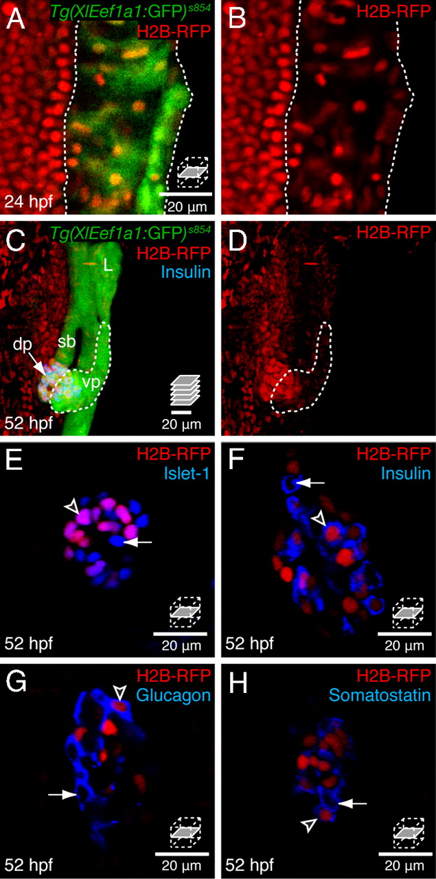
The principal islet contains label-retaining endocrine cells. Embryos were labeled by injection of H2B-RFP mRNA at the one-cell stage. (A and B) Confocal sections through the endoderm at 24 hpf, and (C and D) confocal projections of endodermal tissue at 52 hpf, anterior to the top, dorsal to the left; L, liver, sb, swim bladder, dp, dorsal pancreas, vp, ventral pancreas. Dotted lines delineate Tg(XlEef1a1:GFP)s854 expressing endodermal tissue (A and B) or the ventral pancreatic bud (C and D). (E–H) Confocal sections through the principal islet. (A and B) All cell nuclei are initially labeled with H2B-RFP, including the GFP positive endodermal tissue (dotted outline). (C and D) At 52 hpf, the H2B-RFP label is retained in a dorsal cluster of endodermal cells, some of which coexpress Insulin (blue). Ventral pancreatic tissue (dotted outline) has diluted the H2B-RFP label. (E) All label-retaining endodermal cells coexpress the transcription factor Islet-1, arrowhead. (F–H) A subset of label-retaining endodermal cells express Insulin (F) Glucagon (G) or Somatostatin (H), arrowheads. (E–H) All islets at this stage also contain cells that are H2B-RFP negative but Islet-1 and hormone positive, arrows.