
Fig. 1
- ID
- ZDB-IMAGE-101118-22
- Genes
- Publication
- Schuster et al., 2010 - Glial cell line-derived neurotrophic factor defines the path of developing and regenerating axons in the lateral line system of zebrafish
- All Figures
- Figures for Schuster et al., 2010
|
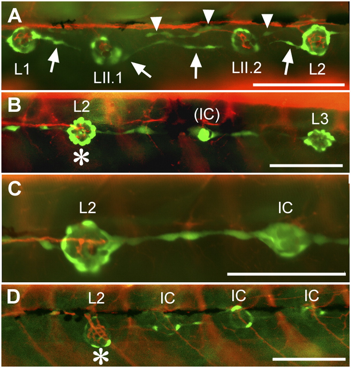
Fig. 1
Innervation defects in Huc:kaede, Et20:gfp early larvae. (A) Normal pattern in a control larva at 4 dpf. Nerve branches extend to and arborize in each neuromast. Interneuromast cells deposited by the primordium between L1 and L2 (arrows) have moved ventrally ahead of the primII-derived neuromasts, LII.1 and LII.2. Interneuromast cells deposited by primII can also be observed close to the myoseptum (arrowheads). (B) Peripheral afferent axons stop at L2 (asterisk) in a 3-dpf gdnf, ret1-MO1 fish. An intercalary neuromast (IC) is beginning to develop between L2 and L3. The red fibers posterior to L2 belong to motor axons innervating the somitic muscles. (C) A higher magnification photograph to show that no nerve branch extends beyond L2, in a 4-dpf double morphant fish. (D) Nerve stop at L2 (asterisk) in a 4-dpf gdnf-MO2 fish. Intercalary neuromasts have formed on all somitic borders posterior to the nerve arrest. Anterior is Left in all panels. (Scale bar, 100 mm.)