
|
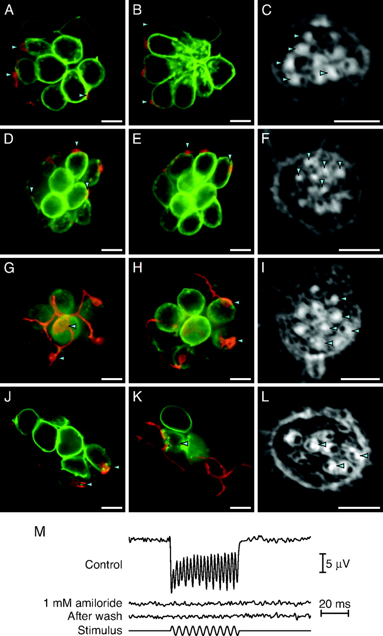
Fig. 2
Stimulus-evoked patterns of synaptic release are not required for polarity choice. (A and B) In an anteroposterior neuromast of a tmie mutant larva, a labeled afferent fiber synapses with five of the ten hair cells. In this and the subsequent morphological illustrations, the two micrographs represent different planes of focus. (C) The hair-bundle polarities of this neuromast reveal that the neuron innervates all five posteriorly polarized hair cells and none of the opposite polarity. (D–F) In a dorsoventral neuromast of a tmie mutant, an afferent neuron innervates only the five ventrally polarized hair cells. (G–I) An afferent fiber in a pcdh15a mutant forms synapses with four of the five anteriorly polarized hair cells but with none of the five cells of the opposite polarity. (J–L) In a neuromast of a larva treated with 1 mM amiloride from 2 dpf to 5 dpf, the labeled fiber forms synapses with only the three anteriorly polarized hair cells. (M) The microphonic potential recorded from a neuromast of a 5-dpf larva under control conditions (Top trace) reveals a response at twice the frequency of the 200-Hz, ±8-μm stimulus (Bottom trace). Stimulation of a neuromast from a sibling maintained for 3 days in 1 mM amiloride reveals no microphonic signal (Second trace). Even after extensive washout of the amiloride, the neuromast fails to respond (Third trace).