
|
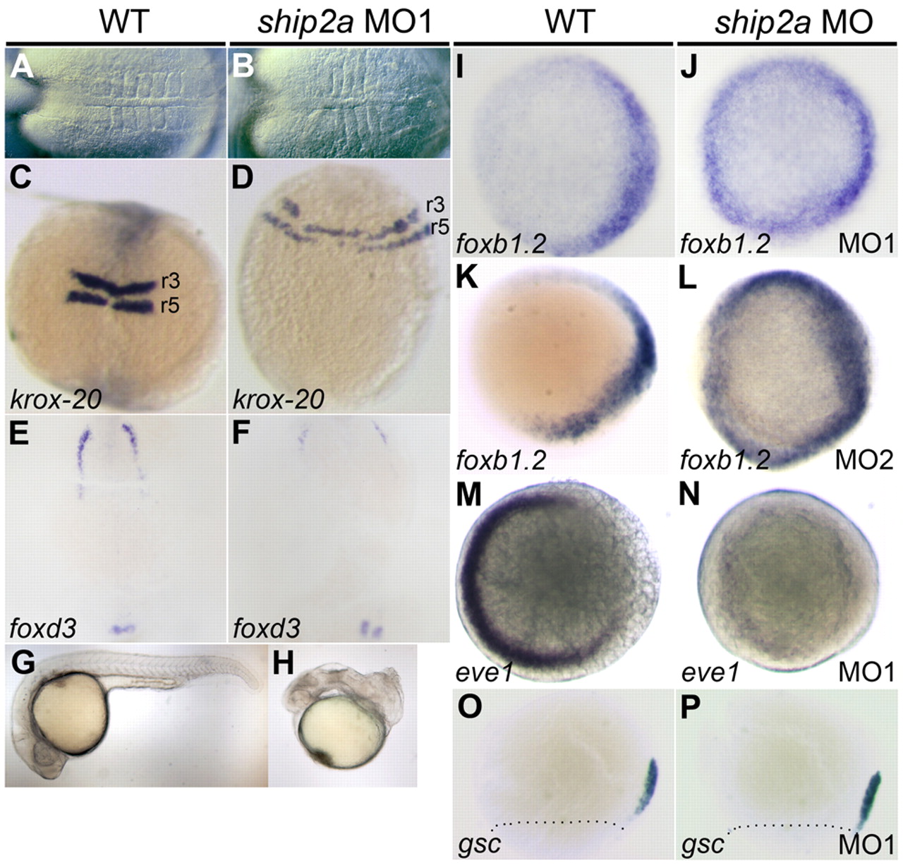
Fig. 2 Knockdown of maternal Ship2a dorsalizes the embryo and affects patterning prior to the onset of gastrulation. (A-F) Lateral expansion of dorsal tissue fates in 5–6-somite stage embryos depleted for Ship2a protein. Maternal ship2a-morphant embryos have a lateral expansion of somites (A,B) and a lateral expansion of krox-20 expression in rhombomeres 3 and 5 (r3 and r5) (C,D) compared with WT controls. (E,F) foxd3 expression at the lateral border of the neural plate is reduced in maternal ship2a-morphant embryos. (G,H) ship2a morphants that survive to 24 hpf appear dorsalized. (I–N) foxb1.2 and eve1 expression at the sphere stage (4 hpf) in WT and maternal ship2a-morphant embryos. (I,K) foxb1.2 is expressed dorsally in WT embryos, and (J,L) its expression is expanded to the ventral side of the embryo in maternal ship2a morphants. (M) eve1 expression is restricted to the ventral side in WT embryos, and (N) ventral eve1 expression is lost in maternal ship2a morphants. (O,P) goosecoid (gsc) expression marking the dorsal organizer at 70% epiboly is unchanged between WT and maternal ship2a-morphant embryos. A and B, dorsal views, anterior to the left; C-F, dorsal views, anterior to top; G and H, lateral views; I-N, animal pole views with dorsal to the right; O and P, lateral views, dorsal to right, markings indicate blastoderm margin.