
Fig. 1
- ID
- ZDB-IMAGE-101104-81
- Publication
- Imrie et al., 2010 - White adipose tissue development in zebrafish is regulated by both developmental time and fish size
- All Figures
- Figures for Imrie et al., 2010
|
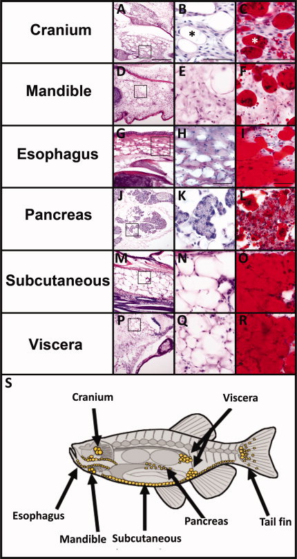
Fig. 1 White adipose tissue (WAT) is located in distinct depots in adult zebrafish. A-R: Serial cryosections from adult (12-18 months old) zebrafish were stained with H&E (A,B,D,E,G,H,J,K,M,N,P,Q) or oil red O (C,F,I,L,O,R) for neutral lipids. The boxed region in the far left panels is magnified in the middle panels. Adipocytes have a unilocular lipid droplet (B,C asterisk) thin cytoplasm and peripheral nucleus (B,C arrow). The pancreatic acinar cells are labeled in K. Scale bar = 200 μm in the right panels and 50 μm in the middle and left panels. S: Diagram of the major sites of WAT in zebrafish adults (yellow circles).