
Fig. 4
- ID
- ZDB-IMAGE-101104-45
- Publication
- Hui et al., 2010 - Cellular response after crush injury in adult zebrafish spinal cord
- All Figures
- Figures for Hui et al., 2010
|
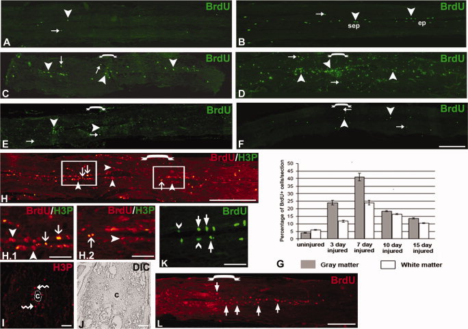
Fig. 4 Time course analysis of injury-induced cell proliferation represented after BrdU incorporation and H3P staining in uninjured and injured spinal cord. A, B: Uninjured cord shows BrdU+ cells are present both in grey matter (▼) and white matter (→) but mostly in ependyma (ep). C: 3dpi cord shows a large increase in BrdU+ cells in both grey (▼) and white (→) matter. D: A section of 7dpi cord shows a very significant increase in number of BrdU+ cells in both grey (▼) and white matter (→), and increased numbers of cells are present at the injury epicenter ([down bracket]) compared to 3dpi cord. E: A 10dpi cord shows incorporation in both white matter (→) and grey matter (π) but a considerable decrease in number of BrdU+ cells compared to 7dpi cord. F: A 15dpi cord section shows fewer BrdU+ cells in grey (▼) and white matter (→) compared to the 10dpi cord and the number is still higher than in the uninjured cord. G: Quantitative representation of BrdU+ cells in both grey and white matter of uninjured and injured cord at different time points. H: A 7dpi cord section shows BrdU+ cells (▼) and many of these are colocalized with H3P (↓) in both the injury epicenter ([down-bracket]) and adjacent part. H.1, H.2: Same 7dpi cord as in H showing adjacent area and injury epicenter, respectively, at high magnification. I, J: Another representative cross-section of 7dpi cord showing distribution of H3P+ cells (squiggly arrow) around the central canal (c) and DIC of same section. K: Ependyma of uninjured cord showing both slow-dividing (thick arrow) and fast-dividing (∨) cells. L: Section of 7dpi cord showing BrdU+ slow-dividing cells (thick arrow) after 48-hr continuous BrdU pulse. Scale bar = 400 μm (A-H), 20 μm (H.1-K), 200 μm (L).