
|
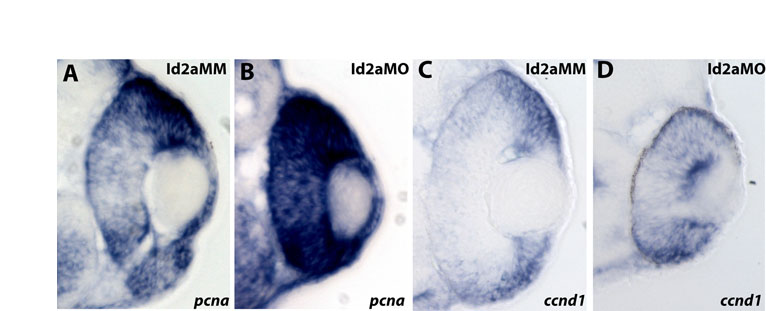
Fig. S4 Loss of Id2a leads to a failure to restrict the expression of pcna and ccnd1 in the developing retina. (A-D) Cryosections reveal the expression domains of pcna at 34 hpf (A,B) and ccnd1 at 48 hpf (C,D) in Id2a-MM retinas and Id2a-MO retinas, respectively. At 34 hpf, the expression of pcna remains high throughout the peripheral margins of the eye, whereas in Id2a-MO retinas expression remains strong throughout the central retina. Similarly, the expression of ccnd1 remains strong at 48 hpf throughout the Id2a-MO retina (D), whereas its expression in the Id2a-MM retina (C) is restricted to the peripheral margins of the eye.