
Fig. 5
- ID
- ZDB-IMAGE-101025-2
- Genes
- Publication
- Larson et al., 2010 - Defective adult oligodendrocyte and Schwann cell development, pigment pattern, and craniofacial morphology in puma mutant zebrafish having an alpha tubulin mutation
- All Figures
- Figures for Larson et al., 2010
|
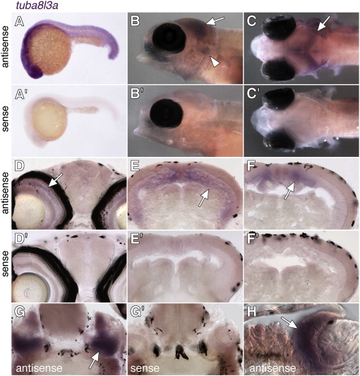
Fig. 5 tuba8l3a is not spatially restricted in the embryo but is expressed most prominently in the central nervous system during post-embryonic development. Shown is staining with antisense and sense probes (diluted to equal concentrations) targeted to the 52 untranslated region of tuba8l3a, (A, A′) Widespread expression of tuba8l3a at 24 hpf. (B, B′) Lateral views of larvae (7.2 SSL) showing tuba8l3a transcript in the brain (arrow) and cranial ganglia (arrowhead). (C, C′) Dorsal views of the same individuals, illustrating tuba8l3a mRNA in the brain (arrow). Larvae in B–C are homozygous nacre mutants that lack otherwise obscuring melanophores due to an autonomously acting mutation in the mitfa transcription factor. Expression in wild-type larvae was indistinguishable from nacre mutants (not shown). (D, D′) tuba8l3a transcript is detectable in the inner nuclear layer of the retina (arrow). (E, E′, F, F′) More posteriorly, tuba8l3a is expressed in the periventricular grey zone of optic tectum. (G, G′, H) tuba8l3a staining (arrow) in the cranial ganglia. Larvae in D–H are 8–9 SSL.
Reprinted from Developmental Biology, 346(2), Larson, T.A., Gordon, T.N., Lau, H.E., and Parichy, D.M., Defective adult oligodendrocyte and Schwann cell development, pigment pattern, and craniofacial morphology in puma mutant zebrafish having an alpha tubulin mutation, 296-309, Copyright (2010) with permission from Elsevier. Full text @ Dev. Biol.