
|
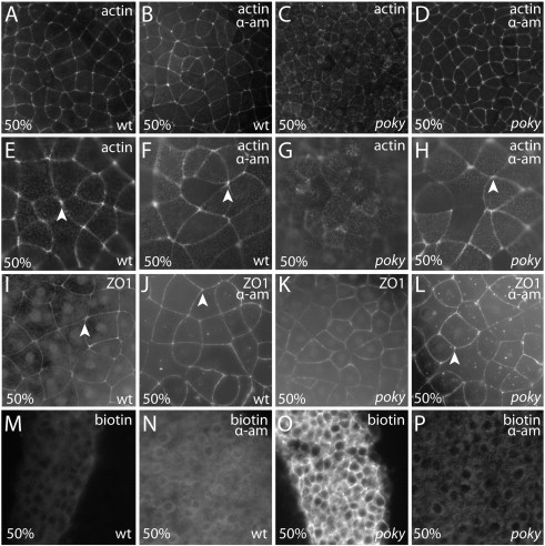
Fig. 6 poky mutant phenotype requires zygotic transcription. (A–H) EVL of phalloidin stained embryos: (A–D) low magnification and (E–H) high magnification. Untreated (A,E) and alpha-amanitin treated (B,F) wild type embryos display similar organization of actin microfilaments with robust localization to cell borders and cell vertices (arrowheads). Untreated poky mutants (C,G) display smaller EVL cells with less robust localization of actin to the EVL cell borders. Alpha-amanitin treated poky mutants (D,H) display a wild type EVL cell morphology, tight actin localization to the cell boundaries, and robust vertex labeling (arrowhead). (I–L) ZO1 localization at the same magnification as E–H. Untreated (I) and alpha-amanitin treated (J) wild type embryos display similar localization of ZO1 to tight junctions. Untreated poky mutants (K) display more EVL cells with less robust localization of ZO1 to cell borders and vertices. Alpha-amanitin treated poky mutants (L) display a wild type EVL cell morphology and tight ZO1 localization to the cell boundaries and robust localization to the vertices (arrowhead). (M,N) Wild type untreated and treated embryos exclude biotin from the DEL. (O) poky mutants do not exclude biotin and show strong interstitial labeling with fluorescent streptavidin. (P) Alpha-amanitin treated poky embryos exclude biotin from the DEL.
Reprinted from Developmental Biology, 346(2), Fukazawa, C., Santiago, C., Park, K.M., Deery, W.J., Canny, S.G., Holterhoff, C.K., and Wagner, D.S., poky/chuk/ikk1 is required for differentiation of the zebrafish embryonic epidermis, 272-283, Copyright (2010) with permission from Elsevier. Full text @ Dev. Biol.