
|
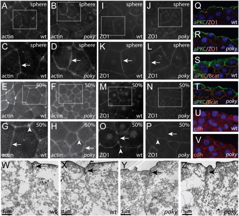
Fig. 3 poky mutant embryos display junction protein localization and apical–basal polarity. Wild type embryos displayed tight localization of actin microfilaments at cell–cell boundaries (arrows) at sphere (A, magnified in C) and robust labeling of cell vertices at 50% epiboly (E magnified in G, arrowheads). poky mutant embryos displayed actin localization to cell–cell boundaries (arrows) at sphere stage (B magnified in D). At 50% epiboly (F magnified in H) the localization to cell–cell boundaries was less robust. Gaps were observed between neighboring cells (arrows) and little labeling was observed at cell vertices (arrowheads). Wild type embryos displayed localization of ZO1 to the tight junction at EVL cell borders at sphere (I magnified in K) and 50% epiboly (M magnified in O) with strong localization to cell–cell boundaries (arrows) and cell vertices (arrowheads). poky mutants also displayed localization at sphere stage (J, magnified in L). By 50% epiboly poky mutant cells were smaller but still displayed localized ZO1 (N magnified in P, arrow), although they lacked cell vertex labeling (arrowhead, N,P). Wild type (Q,S,U) and poky mutant (R,T,V) embryos display apical localization of aPKC (green, Q–T), localization of ZO1 to sites of EVL cell–cell contact (red, Q,R), basolateral localization of β-catenin (red, S,T) and cadherin (U,V). (W–Z) TEM of wild type (W,X) and poky mutant (Y,Z) EVL cells at 30% epiboly (tight junctions, black arrows).
Reprinted from Developmental Biology, 346(2), Fukazawa, C., Santiago, C., Park, K.M., Deery, W.J., Canny, S.G., Holterhoff, C.K., and Wagner, D.S., poky/chuk/ikk1 is required for differentiation of the zebrafish embryonic epidermis, 272-283, Copyright (2010) with permission from Elsevier. Full text @ Dev. Biol.