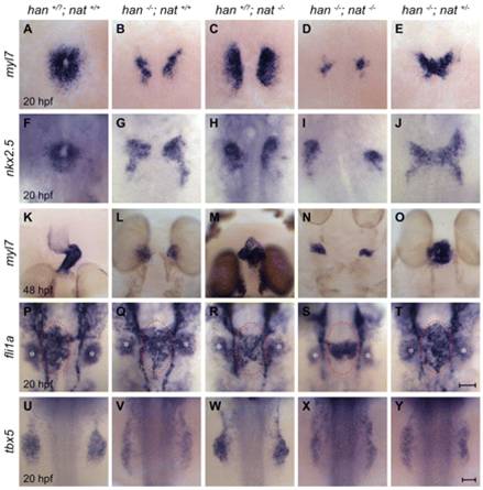
Fig. 3 Reduction of fn1 function rescues cardiac fusion in hand2 mutants. In situ hybridizations for the indicated genes. Dorsal views, anterior up. Zebrafish embryos were genotyped by PCR following imaging, and all phenotypes were found to be consistent; for example, the rescue observed in E was seen in a total of 18 embryos from three independent clutches. (A-J) Cardiac fusion progresses in wild-type and han-/-nat+/- embryos, whereas cardia bifida is present in han, nat and han-/-nat-/- embryos. (K-O) In contrast to the wild-type midline heart, a cardiac cone forms in han-/-nat+/- embryos, a bifurcated heart forms in nat mutants, and cardia bifida is seen in han and han-/-nat-/- embryos. The nat cardiac phenotype varies, ranging from a midline heart to cardia bifida (Trinh and Stainier, 2004). (P-T) Endocardial sheet formation in wild-type and han-/-nat+/- embryos contrasts with the dysmorphic endocardium in han, nat and han-/-nat-/- embryos. Red ovals mark the extent of anterior-posterior spreading of the endocardial sheet in wild-type and han-/-nat+/- embryos; this spreading appears inadequate in han, nat and han-/-nat-/- embryos, either due to morphogenesis or cell number defects. In addition to its endothelial expression, fli1a is expressed in branchial arch mesenchyme (asterisks). (U-Y) The failure of pectoral fin mesenchyme development in han embryos is not modified by altering nat function, in contrast to the normal fin mesenchyme observed in wild-type or nat embryos. Scale bars: 50 μm.
Image
Figure Caption
Acknowledgments
This image is the copyrighted work of the attributed author or publisher, and
ZFIN has permission only to display this image to its users.
Additional permissions should be obtained from the applicable author or publisher of the image.
Full text @ Development

