
Fig. 4
- ID
- ZDB-IMAGE-101011-28
- Antibodies
- Publication
- Zanazzi et al., 2010 - Enrichment and differential targeting of complexins 3 and 4 in ribbon-containing sensory neurons during zebrafish development
- All Figures
- Figures for Zanazzi et al., 2010
|
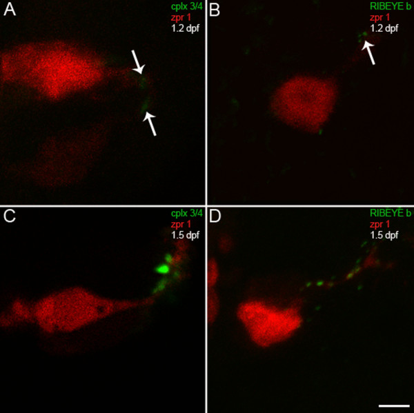
Fig. 4 Complexin 3/4 concentrates in pineal photoreceptor terminals concomitant with RIBEYE b. (A) A high-magnification confocal projection of zpr 1/FRet 43-positive pineal double cone photoreceptors (red) double-stained for complexin 3/4 (green) at 1.2 dpf shows low levels of complexin in photoreceptor terminals (arrows). Ventral is toward the top and lateral is toward the right. (B) A section from a different embryo at 1.2 dpf double-stained with anti-zpr 1/FRet 43 (red) and anti-RIBEYE b (green) shows three small RIBEYE puncta in a pineal photoreceptor terminal (arrow). (C) By 1.5 dpf, complexin 3/4 (green) is highly expressed in neuropil at the lateral border of the pineal organ, in both zpr 1/FRet 43-positive (red) and zpr 1/FRet 43-negative photoreceptor axons and terminals. (D) Several RIBEYE b puncta (green) are present in pineal photoreceptor axons and terminals at 1.5 dpf. Scale bar = 5 μm.