
Fig. S7
- ID
- ZDB-IMAGE-101011-25
- Publication
- Erickson et al., 2010 - Meis1 specifies positional information in the retina and tectum to organize the zebrafish visual system
- All Figures
- Figures for Erickson et al., 2010
|
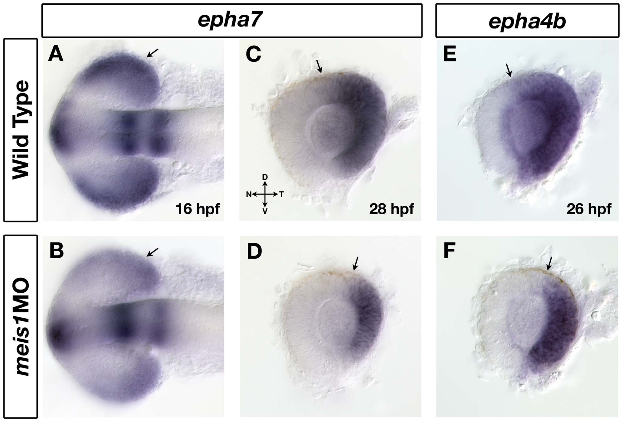
Fig. S7 The temporal expression domains of epha7 and epha4b are reduced in meis1 morphants. (A, B) mRNA in situ hybridization (ISH) for epha7 on wild-type (A) and ,i>meis1 morphant (B) embryos at 16 hpf. Arrows indicate the expression of epha7 in the presumptive temporal retina. Embryos are shown in dorsal view with anterior to the left. (C-F) mRNA ISH for the temporal markers epha7 (C, D) and epha4b (E, F) in dissected, flat-mounted eyes from 26- to 28-hpf wild-type and meis1 morphant embryos. Arrows indicate the dorsal extent of gene expression. Representative dissected eyes are shown. Legend for retinal axial orientation: D, dorsal; V, ventral; N, nasal; T, temporal.