
IMAGE
Fig. 7
- ID
- ZDB-IMAGE-101008-13
- Publication
- Imai et al., 2010 - The ubiquitin proteasome system is required for cell proliferation of the lens epithelium and for differentiation of lens fiber cells in zebrafish
- All Figures
- Figures for Imai et al., 2010
Image
|
Figure Caption
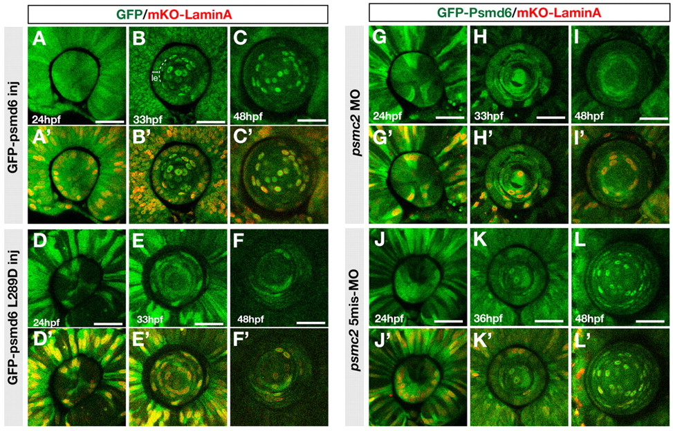
Fig. 7 Localization of GFP-tagged Psmd6 in lens fiber cell nuclei. (A-F′) Localization of GFP-tagged Psmd6 (green) (A-C) and GFP-tagged Psmd6 carrying the L289D mutation (green) (D-F) in wild-type zebrafish lens of the indicated stages. Nuclear membranes are visualized with mKO-Lamin A (red) (A′-F′). le, lens epithelial region. (G-L′) Localization of GFP-tagged Psmd6 in psmc2 morphant (G-I) and psmc2 five-mismatch morphant (J-L) lens. Nuclear membranes are visualized with mKO-Lamin A (red) (G′-L′). All panels are anterior views of the lens sphere. Scale bars: 50 µm.
Acknowledgments
This image is the copyrighted work of the attributed author or publisher, and
ZFIN has permission only to display this image to its users.
Additional permissions should be obtained from the applicable author or publisher of the image.
Full text @ Development