
Fig. 9
- ID
- ZDB-IMAGE-101004-38
- Publication
- Kelsh et al., 1996 - Zebrafish pigmentation mutations and the processes of neural crest development
- All Figures
- Figures for Kelsh et al., 1996
|
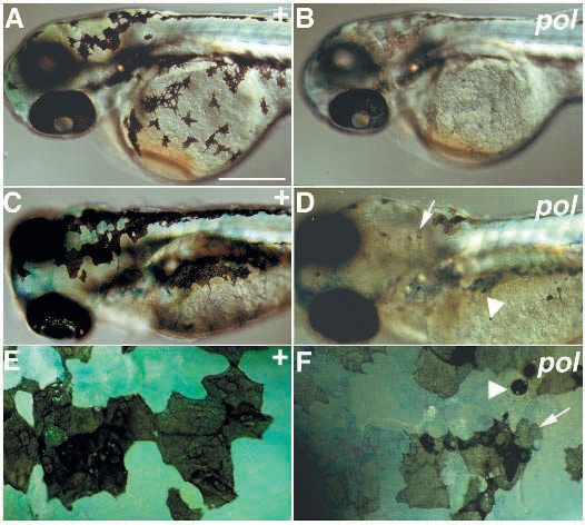
Fig. 9 Phenotype of melanophore degeneration and pale xanthophore mutants (Class VI.D). Wild-type siblings are compared with homozygous mutants for pol on day 2 (A,B) and day 5 (C-F). Melanophores on the third day are of normal size, but are very pale (pigmented retinal epithelium is also pale) (B). By the sixth day many melanophores are abnormal in shape (arrow, D,F) and some are small and spot-like (arrowhead, D,F). Xanthophore pigmentation is paler (B,D,F): note that blue colour of xanthophores is due to their taking up methylene blue from the medium. A-D are dorsolateral views; E-F are dorsal views of the head dorsal stripe. Scale bars, 250 μm (A,B), 175 μm (C,D) and 45 μm (E,F).