
IMAGE
Fig. 3
- ID
- ZDB-IMAGE-101004-3
- Antibodies
- Publication
- Kelsh et al., 2000 - The zebrafish colourless gene regulates development of non-ectomesenchymal neural crest derivatives
- All Figures
- Figures for Kelsh et al., 2000
Image
|
Figure Caption
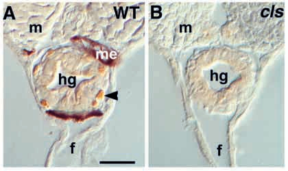
Fig. 3 Enteric neurons are extremely reduced in cls- larvae. Transverse sections of posterior trunk of (A) wild-type larva at 7 dpf stained for the anti-Hu epitope reveal numerous Hu-positive neurons (arrowhead) around the hindgut (hg). A sibling cls- larva (B) lacks these cells. In whole-mount hindguts, dissected at 5 dpf, cls- larvae have only about 13% as many enteric neurons (28.4±10.9; n=7), compared to wild types (212±20.9; n=3). f, ventral fin; m, myotome; me, melanophore. Scale bar, 35 μm.
Figure Data
Acknowledgments
This image is the copyrighted work of the attributed author or publisher, and
ZFIN has permission only to display this image to its users.
Additional permissions should be obtained from the applicable author or publisher of the image.
Full text @ Development