
Fig. 2
- ID
- ZDB-IMAGE-101004-16
- Publication
- Elworthy et al., 2003 - Transcriptional regulation of mitfa accounts for the sox10 requirement in zebrafish melanophore development
- All Figures
- Figures for Elworthy et al., 2003
|
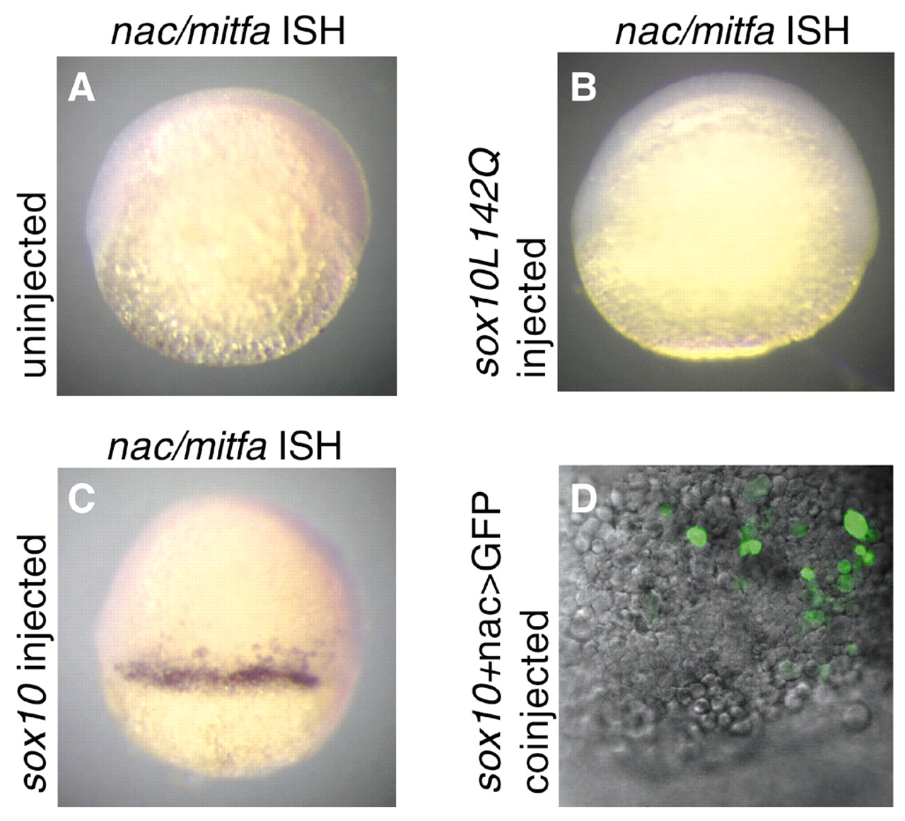
Fig. 2 Precocious nac/mitfa expression in 6 h.p.f embryos following injection with cls/sox10 RNA. Lateral views of uninjected (A), cls/sox10L142Q RNA injected (250 pg per embryo; B) and cls/sox10 RNA injected (250 pg per embryo; C) 6 hpf embryos following in situ hybridization with a nac/mitfa probe. Spots and/or patches of nac/mitfa expression were detected in 39% of cls/sox10 RNA injected embryos (n=136) but not in any of the uninjected embryos (n=58) nor in any of the cls/sox10L142Q RNA injected embryos (n=92). (D) Superimposed fluorescent confocal and DIC images of an animal/lateral view of a 6 hpf embryo coinjected with cls/sox10 RNA (250 pg per embryo) and nac>GFP reporter plasmid (150 pg per embryo) show cells with GFP fluorescence. GFP fluorescence was observed in 75% (n=224) of embryos coinjected with cls/sox10 RNA and nac>GFP.