
Fig. 4
- ID
- ZDB-IMAGE-100913-36
- Genes
- Publication
- Goh et al., 2010 - The RhoA GEF Syx is a target of Rnd3 and regulated via a Raf1-like ubiquitin-related domain
- All Figures
- Figures for Goh et al., 2010
|
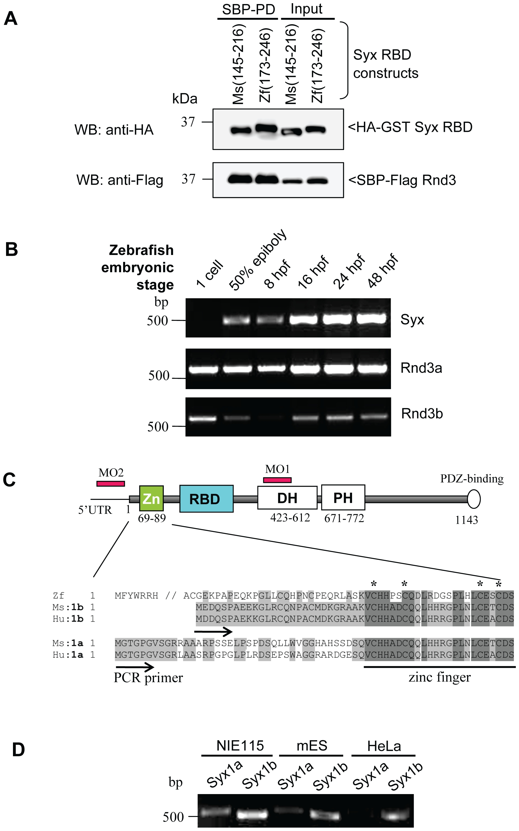
Fig. 4 Zebrafish Syx is closely related to mammalian Syx1b and expressed from epibolic stages.
(A) Zebrafish Syx interacts with mouse Rnd3 via its RBD domain (residues 173–246). (B) Expression of Syx and Rnds mRNA. RT-PCR analysis showing mRNA transcript profiles of zebrafish Syx, Rnd3a and Rnd3b at different developmental stages indicated. Primers to Syx (nucleotide 2640–3120) (XM001923778), Rnd3a (nucleotide 1–732) (BC059452) and Rnd3b (nucleotide 1–672) (BC076009) were used. (C) Schematic representation of zebrafish Syx, including the conserved RBD, DH/PH domains and PDZ-binding motif. The N-terminal region of zebrafish Syx resembles more closely to mammalian Syx1b than the published Syx1a. Conserved residues are in light grey and identical residues in dark grey. The zinc finger motif is underlined and the conserved cysteine residues marked (*). Database numbers for zebrafish Syx (XM_686228.1), mouse Syx1b (B1AS67), human Syx1b (094827), mouse Syx1a (AAU04953) and human Syx1a (AK299523) are in brackets. (D) RT-PCR revelas that Syx 1b is more highly expressed in mouse NIE115, ES and human HeLa cells. RT-PCR from alternative start sites to a common downstream primer (nucleotide 495).