
Fig. 1
- ID
- ZDB-IMAGE-100913-33
- Publication
- Goh et al., 2010 - The RhoA GEF Syx is a target of Rnd3 and regulated via a Raf1-like ubiquitin-related domain
- All Figures
- Figures for Goh et al., 2010
|
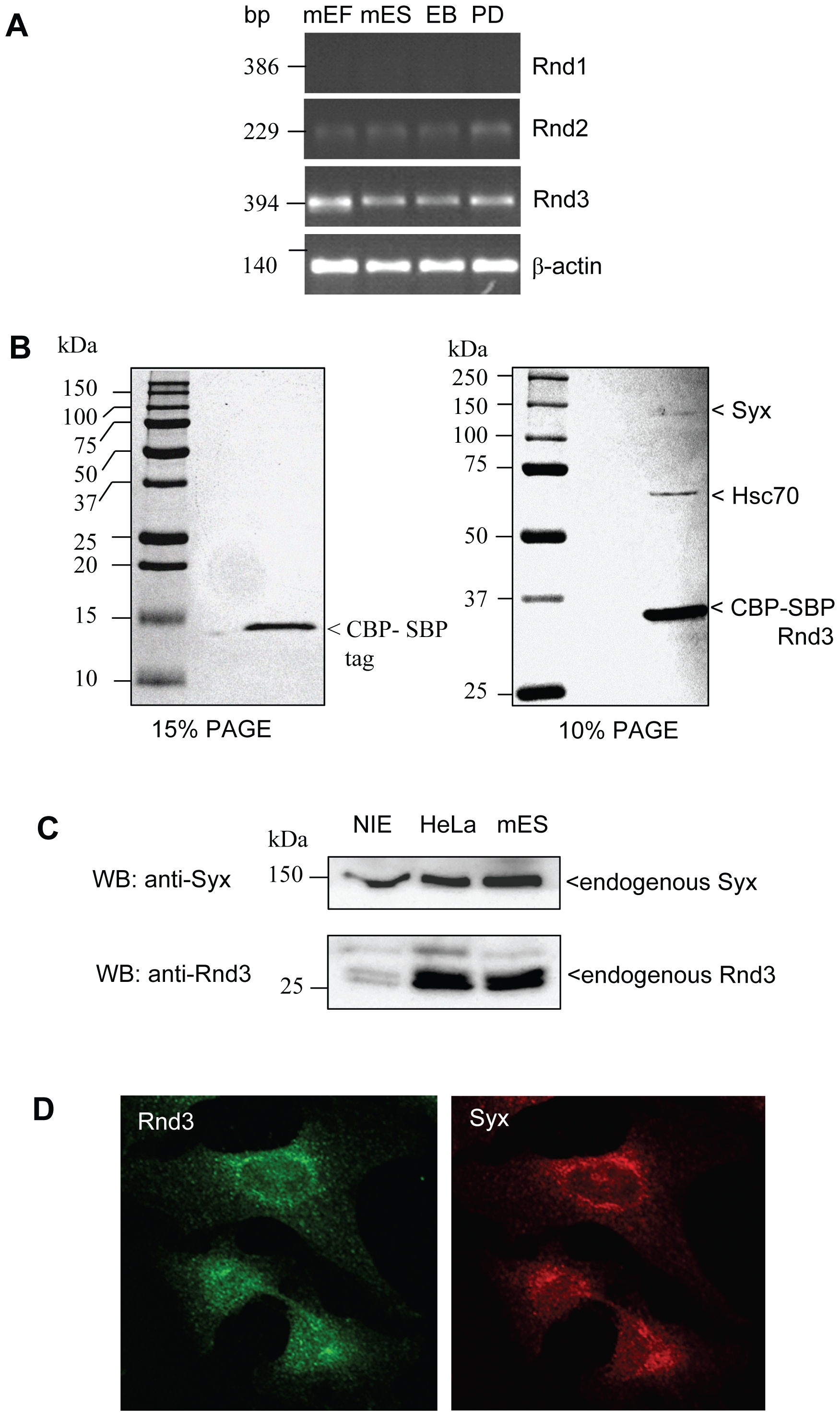
Fig. 1 Rnd3 associates with Syx in mouse embryonic stem cells.
(A) Expression analysis of Rnds evaluated by RT-PCR from mES cells, mouse embryonic fibroblast (mEF) cells, embryoid bodies (EB) or plated differentiated mES cells (PD). (B) Identification of Rnd3-associated proteins. Rnd3 TAP-tagged proteins from transiently transfected mES cells using CBP-SBP vector (see methods). The purified proteins by coomassie staining is shown with identification by LC-MS analysis. (C) Expression levels of Rnd3 and Syx in NIE-115, HeLa and mES cells. Cell lysates were resolved by SDS-PAGE and immunoblotted with anti-Rnd3 and anti-Syx. (D) Endogenous localizations of Rnd3 and Syx in HeLa cells immuno-labeled with anti-Syx and Rnd3 antibodies. Rnd3 and Syx are both localized near the perinuclear region. Bar = 10μM.