
Fig. S1
- ID
- ZDB-IMAGE-100903-58
- Publication
- Wang et al., 2010 - Moesin1 and Ve-cadherin are required in endothelial cells during in vivo tubulogenesis
- All Figures
- Figures for Wang et al., 2010
|
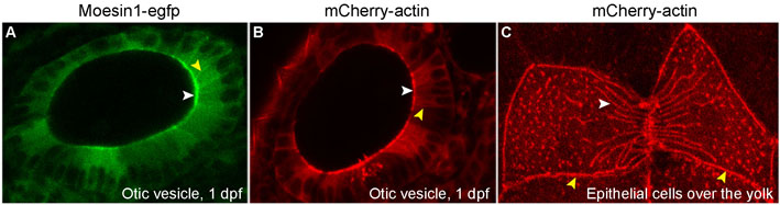
Fig. S1 Moesin1-EGFP and mCherry-β-Actin fusion proteins are localized to the apical membranes and cellular junctions in epithelial cells. RNA encoding Moesin1-EGFP and mCherry-β-Actin was injected into 1-cell stage zebrafish embryos. Localization of the fusion proteins was followed in living embryos. (A,B) Moesin1-EGFP and mCherry-β-Actin both localize to the apical membranes (white arrowheads) and cellular junctions (yellow arrowheads) in epithelial cells in the otic vesicle. (C) mCherry-β-Actin fusion proteins incorporate into the presumptive Actin fibers (white arrowhead) and localize to the presumptive cellular junctions (yellow arrowheads) in epithelial cells in the periderm.