
Fig. 4
- ID
- ZDB-IMAGE-100903-22
- Publication
- Jeong et al., 2010 - Inhibition of Plk1 induces mitotic infidelity and embryonic growth defects in developing zebrafish embryos
- All Figures
- Figures for Jeong et al., 2010
|
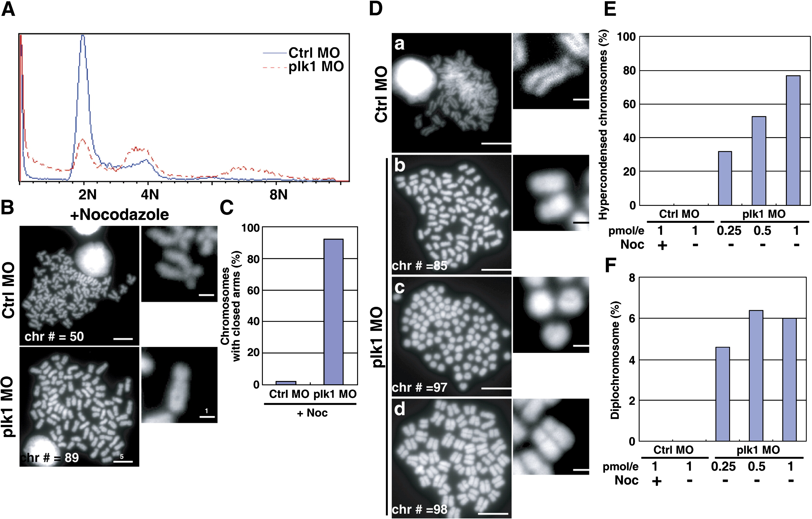
Fig. 4 Severe chromosome abnormalities in plk1 morphant embryos. (A) Embryos were injected with 0.25 pmol of control or plk1 ATG MO and single cell suspensions were prepared at 28 hpf. DNA contents were measured by Propidium Iodide (PI) staining and flow cytometry. (B) Metaphase chromosome spreads from control (Ctrl MO) or Plk1 morphants (Plk1 MO). Chromosome numbers (Chr #) and scale bars are indicated. Enlarged images are shown at the right. (C) Chromosomes with closed arms were counted in control or plk1 morphants and compared and represented as percentages in a bar graph. (D) Representative chromosome spreads prepared without nocodazole treatment. Scale bar, 5 μm. (E) The ratio of hypercondensed chromosomes (Dc), scored from 150 metaphase spreads of 20 embryos each. Data from control morphants treated with nocodazole were included for control. (F) The presence of diplochromosomes (Dd) in plk1 morphants.
Reprinted from Developmental Biology, 345(1), Jeong, K., Jeong, J.Y., Lee, H.O., Choi, E., and Lee, H., Inhibition of Plk1 induces mitotic infidelity and embryonic growth defects in developing zebrafish embryos, 34-48, Copyright (2010) with permission from Elsevier. Full text @ Dev. Biol.