
Fig. 7
- ID
- ZDB-IMAGE-100816-35
- Publication
- Pittman et al., 2010 - nev (cyfip2) Is required for retinal lamination and axon guidance in the zebrafish retinotectal system
- All Figures
- Figures for Pittman et al., 2010
|
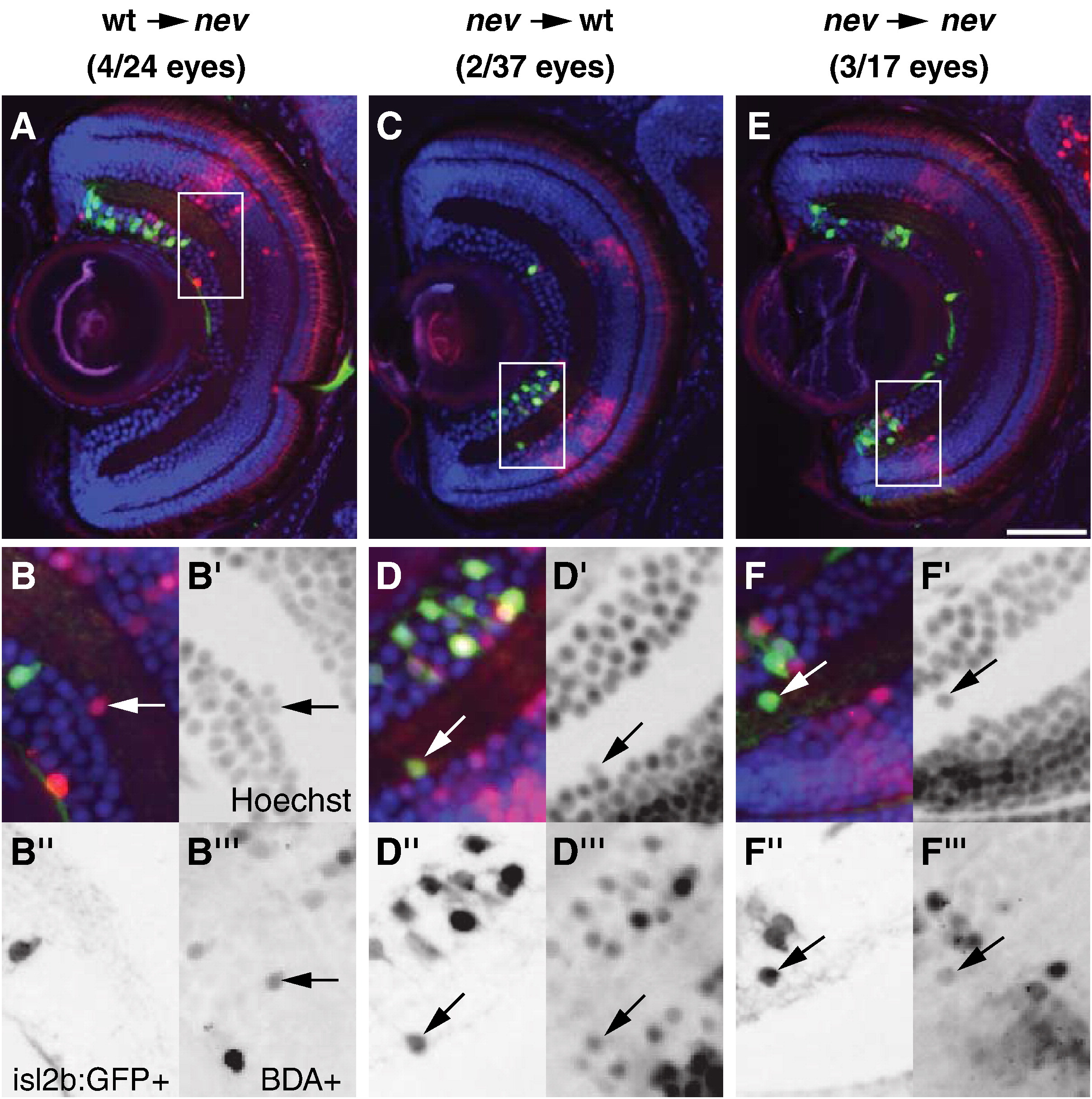
Fig. 7 cyfip2 acts both cell autonomously and cell nonautonomously in lamination. Representative coronal sections through a nev eye with WT donor cells (A–B″′), a WT eye with nev donor cells (C–D″′), and a nev eye with nev donor cells (E-F″′). (A, C, E) Hoechst 33342 stain (blue), biotin dextran-positive (BDA+) donor cells (red), and isl2b:GFP+ donor RGCs (green). Insets are shown magnified below. (B–B″′) WT donor cells in a nev host are misplaced in the IPL, showing nev can act cell nonautonomously. Arrows show a BDA+ donor cell next to a BDA cell in the IPL. (D–D″′) nev donor cells in a WT host are misplaced in the IPL, showing that nev can also act cell autonomously. Arrows show a BDA+/isl2b:GFP+ cell in the IPL. (F–F″′) Control transplants show nev donor cells in a nev host that are misplaced in the IPL. Arrow shows a BDA+/isl2b:GFP+ cell misplaced in the IPL. Scale bar = 50 μm.
Reprinted from Developmental Biology, 344(2), Pittman, A.J., Gaynes, J.A., and Chien, C.B., nev (cyfip2) Is required for retinal lamination and axon guidance in the zebrafish retinotectal system, 784-794, Copyright (2010) with permission from Elsevier. Full text @ Dev. Biol.