
Fig. 2
- ID
- ZDB-IMAGE-100816-30
- Publication
- Pittman et al., 2010 - nev (cyfip2) Is required for retinal lamination and axon guidance in the zebrafish retinotectal system
- All Figures
- Figures for Pittman et al., 2010
|
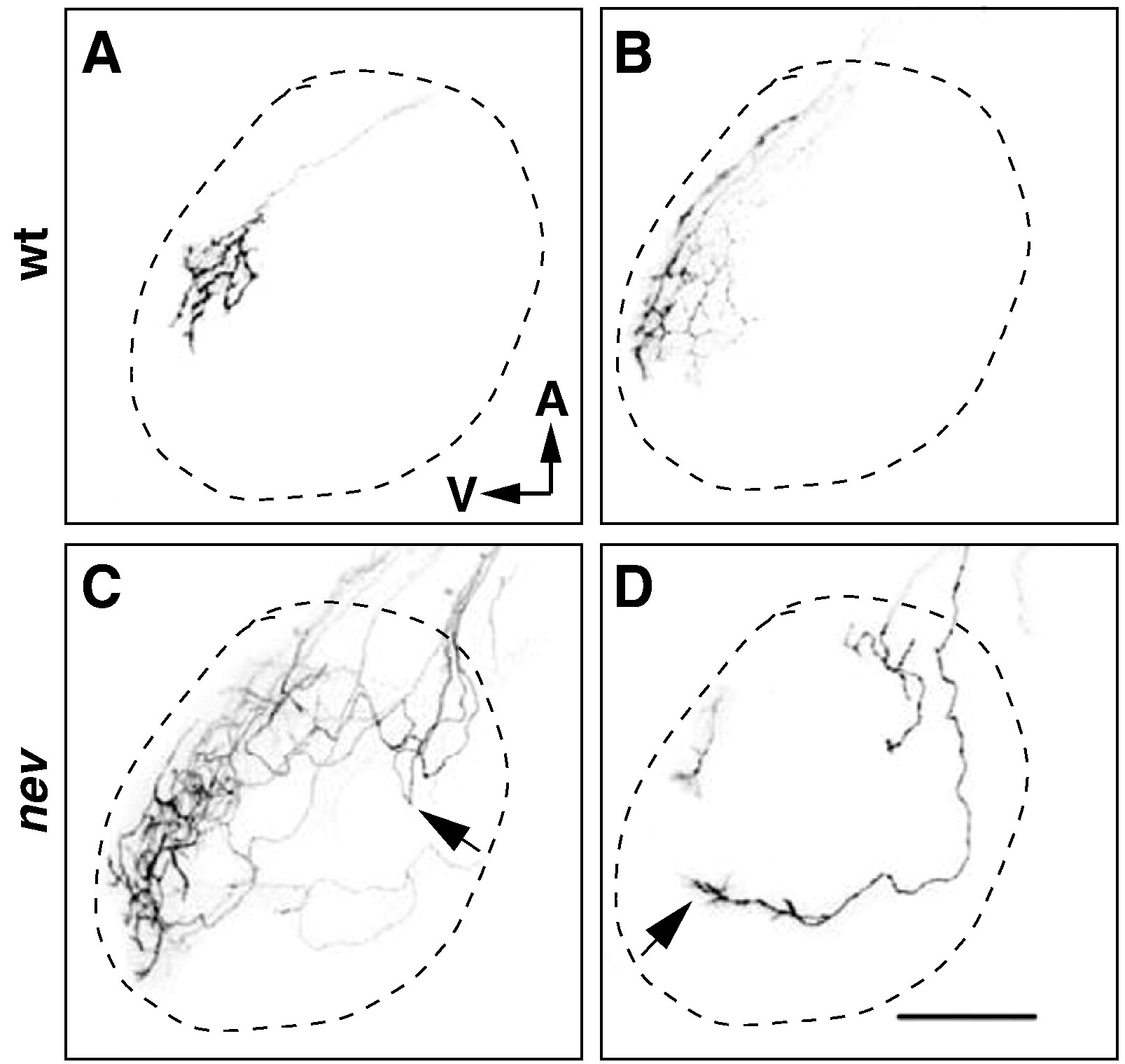
Fig. 2 Dorsonasal axons take circuitous routes on the tectum in nev. (A–D) Confocal images of the left optic tectum after DiI labeling of a few RGCs in the dorsal retina of wt (A and B) or nev (C and D) at 5dpf. Dashed line indicates approximate outline of tectum. In WT, dorsonasal axons enter the tectum through the ventral branch of the optic tract, project directly to their topographic target and arborize (A and B). In nev, dorsonasal axons that enter through the dorsal branch of the optic tract meander around the dorsal tectum, sometimes turning back anteriorly after entering the tectum (arrow in C) but appear to orient toward the ventral tectum (arrow in D). Scale bar in D = 50 μm.
Reprinted from Developmental Biology, 344(2), Pittman, A.J., Gaynes, J.A., and Chien, C.B., nev (cyfip2) Is required for retinal lamination and axon guidance in the zebrafish retinotectal system, 784-794, Copyright (2010) with permission from Elsevier. Full text @ Dev. Biol.