
Fig. S4
- ID
- ZDB-IMAGE-100816-28
- Publication
- Rhodes et al., 2010 - Positive regulation of c-Myc by cohesin is direct, and evolutionarily conserved
- All Figures
- Figures for Rhodes et al., 2010
|
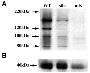
Fig. S4 Validation of the zebrafish-specific Rad21 antibody. Protein lysates were prepared from 20 wild type (WT), sibling (sibs) and rad21nz171 homozygous mutant (mts) embryos at 27 h.p.f. A, Immunoblot showing Rad21 protein. B, α-tubulin loading control. Although there is less total protein in the mutant sample, the Rad21 protein is considerably more reduced in rad21nz171 mutants than can be expected due to loading discrepancy alone. As expected, siblings, comprising a mix of wild type and rad21nz171 heterozygotes, show intermediate levels of Rad21 protein. The band pattern detected, with the major band above 130kDa, is similar to that detected with commercial anti-human Rad21 antibodies.
Reprinted from Developmental Biology, 344(2), Rhodes, J.M., Bentley, F.K., Print, C.G., Dorsett, D., Misulovin, Z., Dickinson, E.J., Crosier, K.E., Crosier, P.S., and Horsfield, J.A., Positive regulation of c-Myc by cohesin is direct, and evolutionarily conserved, 637-649, Copyright (2010) with permission from Elsevier. Full text @ Dev. Biol.