
Fig. 2
- ID
- ZDB-IMAGE-100809-2
- Publication
- Schröter et al., 2010 - Segment Number and Axial Identity in a Segmentation Clock Period Mutant
- All Figures
- Figures for Schröter et al., 2010
|
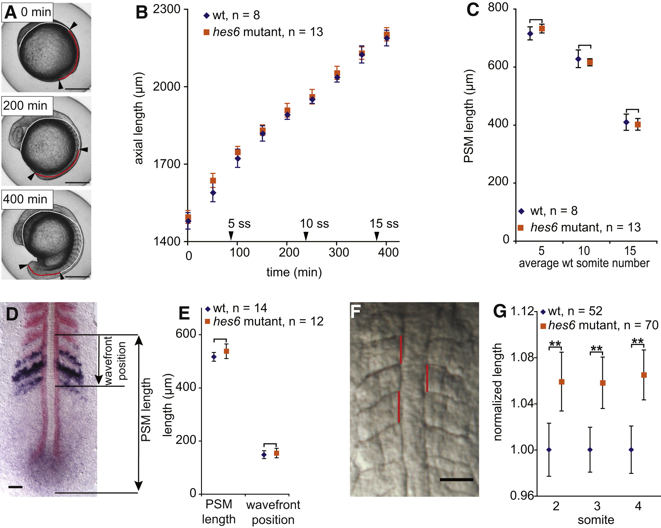
Fig. 2 Unchanged Axial Elongation and Longer Somites in hes6 Mutant Embryos
(A) Total axial length was measured from stills of time-lapse movies by drawing a line (red plus white) from the anterior to the posterior end of the embryo, following the yolk and the dorsal aspect of the paraxial mesoderm. PSM length was measured along the same line (red part) as the distance between the most recently formed somite boundary (arrowhead) and the posterior end of the mesoderm (arrowhead). Three representative stages are shown. The scale bar represents 0.3 mm.
(B) Axial length as measured in (A) is indistinguishable between wild-type and hes6 mutants throughout trunk somitogenesis. Arrowheads on the x axis indicate when wild-type embryos reached the five-, ten-, and 15-somite stage.
(C) PSM length measured as in (A) (red line) is indistinguishable between wild-type and hes6 mutants at three stages during trunk somitogenesis. Measurements were taken at simultaneous time points in all samples; the x axis label indicates average number of somites in wild-type embryos at the respective time point.
(D) PSM length and wavefront position measurement from a fixed PSM stained with myoD (red) to label formed somites and mespb (dark blue) to indicate the wavefront position. Embryo is at the ten-somite stage, flat mount, anterior to the top. The scale bar represents 50 μm.
(E) PSM length and distance of the wavefront from the most recently formed somite boundary as indicated in (D) are indistinguishable between wild-type and hes6 mutant embryos. Genotypes were determined by the presence or absence of hes6 in situ signal (blue staining in the tailbud in D).
One representative experiment from three independent trials is shown in (B) and (C) and from two independent trials in (E). In no case was a difference between wild-type and mutant embryos observed.
(F) Anteroposterior length of somites two to four was measured by drawing a straight line (red) connecting the contact points of the rostral and caudal somite boundaries with the notochord. The scale bar represents 50 μm.
(G) Mean anteroposterior length of somites two to four. Data are pooled from four independent experiments by normalizing mean length of wild-type somites to 1. Somites in hes6 mutants are approximately 6%–7% longer than in their wild-type siblings.
Data in (B), (C), (E), and (G) are given as mean ± 95% CI; **p ≤ 0.001, Student′s t test.