
IMAGE
Fig. S4
- ID
- ZDB-IMAGE-100806-98
- Publication
- Curran et al., 2010 - Interplay between Foxd3 and Mitf regulates cell fate plasticity in the zebrafish neural crest
- All Figures
- Figures for Curran et al., 2010
Image
|
Figure Caption
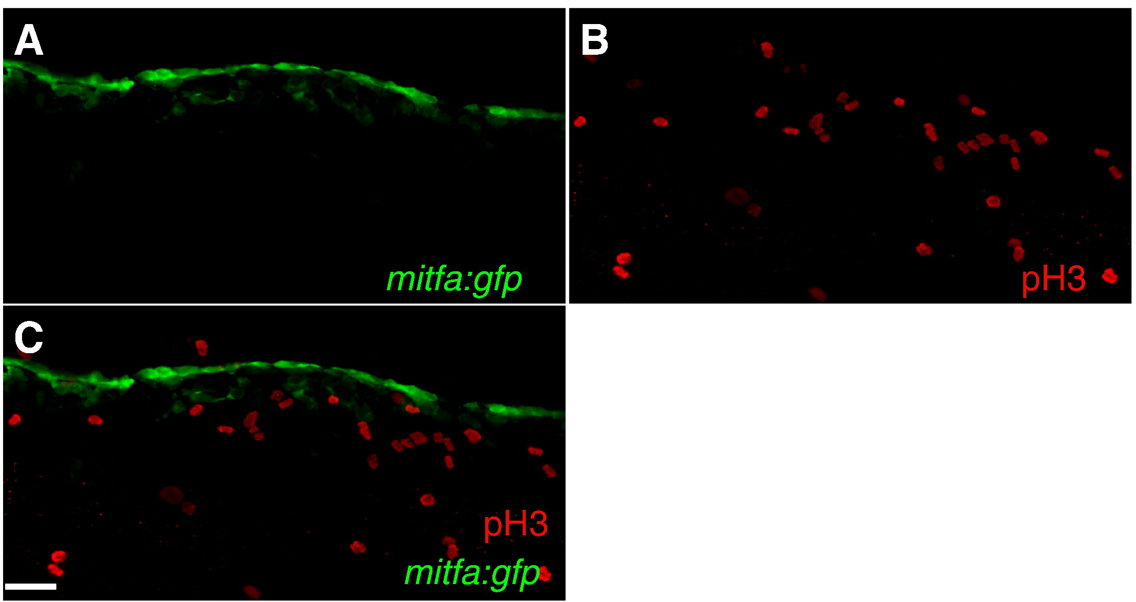
Fig. S4 mitfa:gfp cells are post-mitotic at 24 hpf (A–C) Confocal images collected from lateral aspect of anterior tail region of 24 hpf wild-type zebrafish, anterior left, 20x. (A) mitfa:gfp (B) phosphohistone H3. (C) Color merged: green: mitfa:gfp, red: phosphohistone H3. Scale bars: (A–C) 25 μm.
Acknowledgments
This image is the copyrighted work of the attributed author or publisher, and
ZFIN has permission only to display this image to its users.
Additional permissions should be obtained from the applicable author or publisher of the image.
Reprinted from Developmental Biology, 344(1), Curran, K., Lister, J.A., Kunkel, G.R., Prendergast, A., Parichy, D.M., and Raible, D.W., Interplay between Foxd3 and Mitf regulates cell fate plasticity in the zebrafish neural crest, 107-118, Copyright (2010) with permission from Elsevier. Full text @ Dev. Biol.