
|
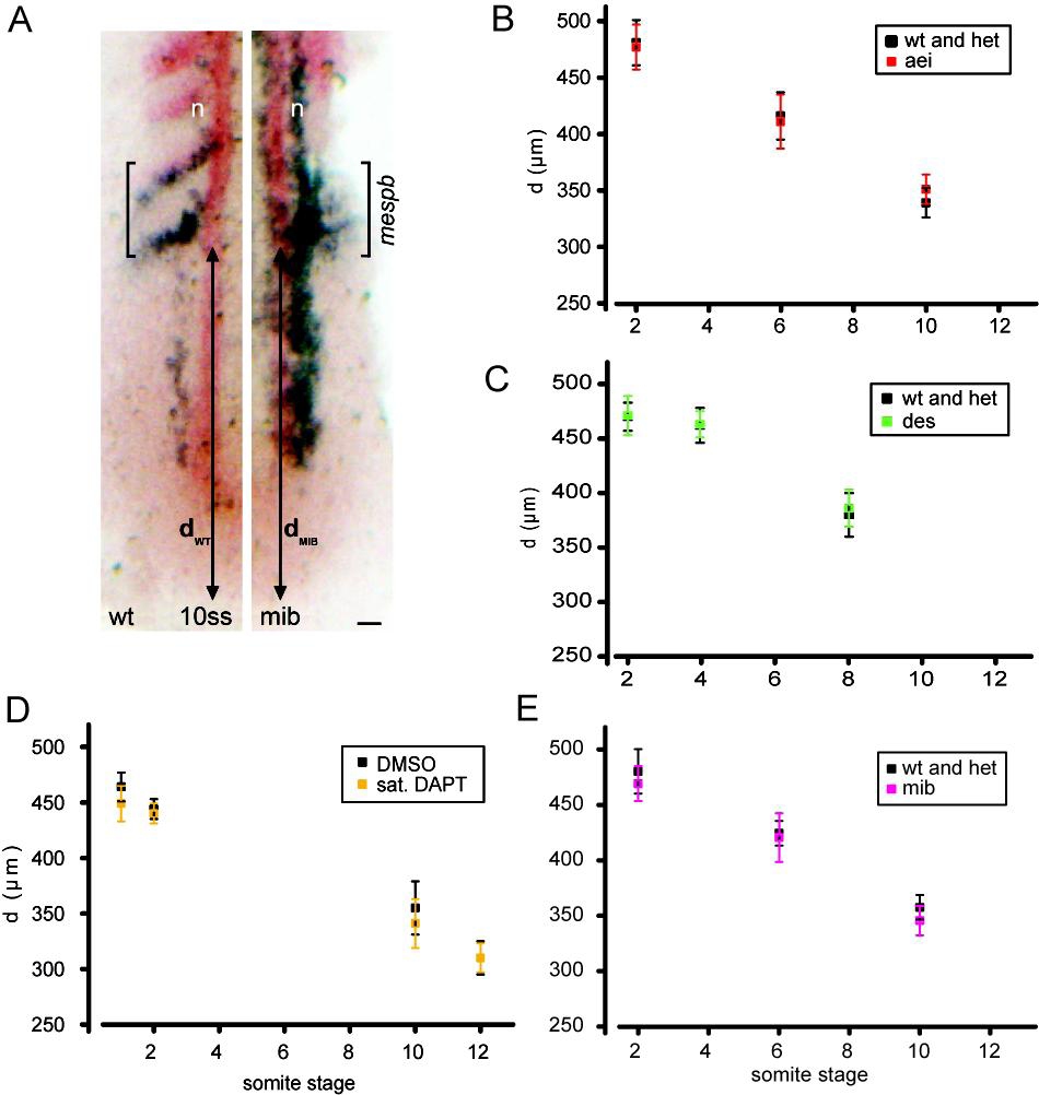
Fig. S3
Position of arrest front is unchanged within the PSM of Delta-Notch mutants and DAPT-treated embryos
(A) In situ hybridization for mespb (blue) and isl1 (blue; expressed in interneurons and Rohon-Beard neurons, n) and myoD (red) in the PSM of wildtype/heterozygous and mib embryos. Bracket: expression pattern of mespb, striped in wildtype and in a salt-and-pepper pattern in mib. Double-headed arrow: d, distance between the posterior boundary of mespb expression domain and the posterior tip of the embryo. Dorsal view with anterior to the top. Scale bar = 25 μm. (B-E) Plots of d (mean ± 95% CI) vs. developmental stage in different experimental conditions, 10 ≤ n ≤ 59 (mean = 22.5) embryos from at least two independent trials for each data point (Table S1). Each experimental condition had three trials for at least one of the data points. Somite stages are given for the wildtpye siblings or clutch mates. Values of arrest front position for control and mutant or DAPT-treated embryos were not significantly different (p > 0.01 in all cases as assessed by Student’s t-test) at any time point or for any of the experimental conditions.