
|
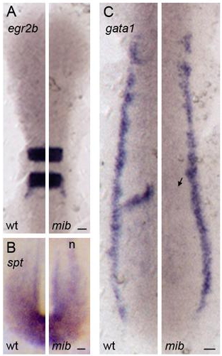
Fig. S1
Expression of mesoderm and ectoderm differentiation markers in mib embryos
In situ hybridization with ectoderm (A) and mesoderm (B, C) markers in the PSM of wildtype/heterozygous and mib embryos at the 10 somite stage. Elevated expression of isl1 in (B) in interneurons and Rohon-Beard neurons (n, out of focal plane) and reduced expression of mespa (arrow in C) is observed in mib embryos, either of which was used to distinguish wildtype/heterozygous from mib embryos. Dorsal view with anterior to the top. Expression patterns were assessed in at least five embryos per experimental condition. Scale bars = 25 μm.