
IMAGE
Fig. 7
- ID
- ZDB-IMAGE-100806-163
- Antibodies
- Publication
- Francescatto et al., 2010 - The activation of membrane targeted CaMK-II in the zebrafish Kupffer's vesicle is required for left-right asymmetry
- All Figures
- Figures for Francescatto et al., 2010
Image
|
Figure Caption
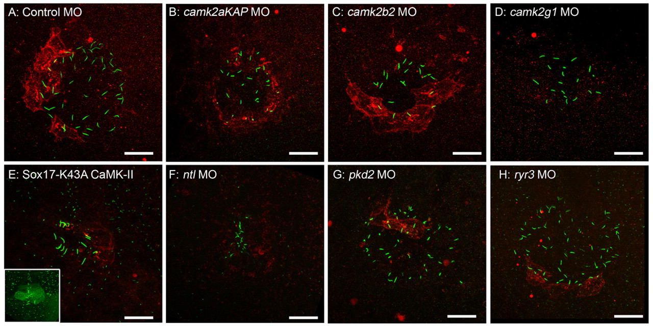
Fig. 7 CaMK-II activation is disrupted in morphants. Confocal immunofluorescent projections of P-T287 CaMK-II (red) and cilia (green) in embryos at the 12-somite stage after injection with (A) 5 ng mismatch (control), (B) 1.5 ng camk2aKAP, (C) 1.5 ng camk2b2, (D) 1.25 ng camk2g1 MO, (E) 150 ng Sox17-K43A GFP-CaMK-II cDNA (inset is green fluorescence of cilia and GFP-CaMK-II around the KV), (F) 4 ng ntl MO, (G) 4 ng pkd2 MO or (H) 4 ng ryr3 MO. Scale bar: 10 μm.
Figure Data
Acknowledgments
This image is the copyrighted work of the attributed author or publisher, and
ZFIN has permission only to display this image to its users.
Additional permissions should be obtained from the applicable author or publisher of the image.
Full text @ Development