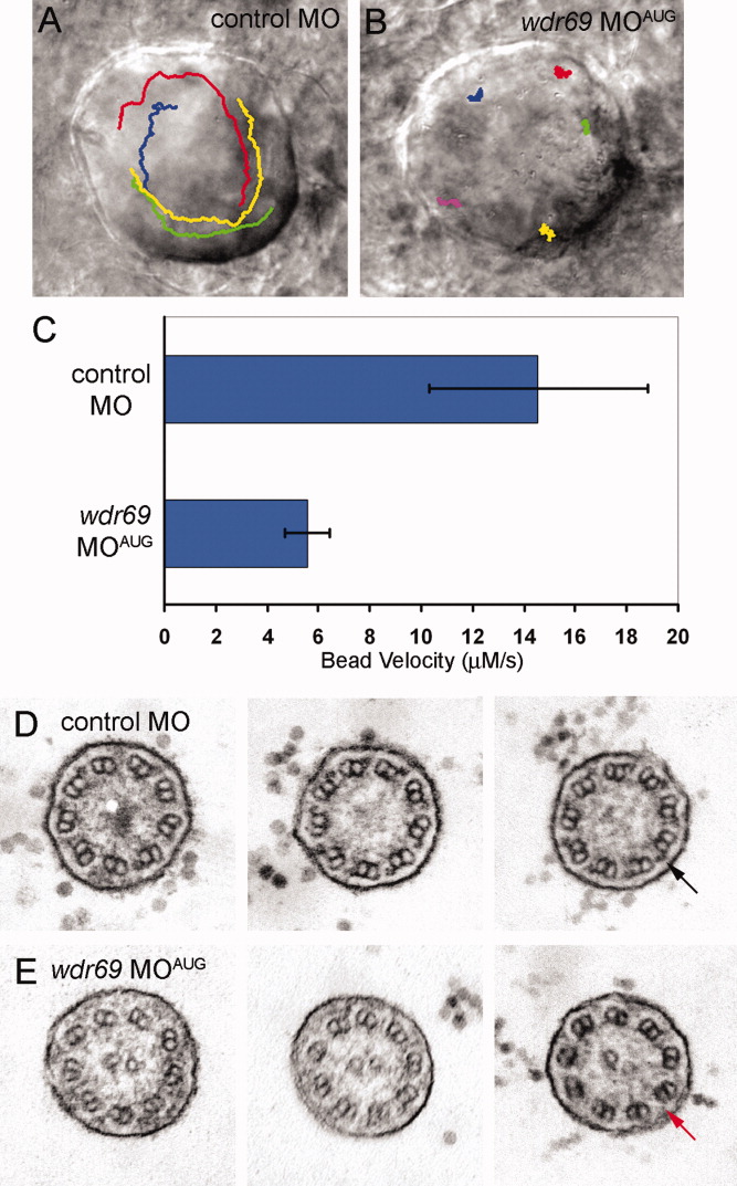
Fig. 5 Wdr69 is needed for ciliary motility and outer arm dynein assembly. A,B: Tracking software was used to follow the flow of fluorescent beads injected into Kupffer′s vesicle (KV) of live embryos. Tracks are superimposed on images of KV. A: In control morphants, beads flowed in a circular counterclockwise direction. B: This directional flow was lost in wdr69 MOAUG morphants, where beads bounced around randomly. C: Velocity of bead movement was significantly reduced in wdr69 knockdown embryos (n = 25 beads from 5 embryos) relative to controls (n = 28 beads from 6 embryos). Error bars=one standard deviation. D,E: Electron microscopy of KV cilia. D: In control MO injected embryos, typical outer dynein arms (arrow) were present on all nine outer doublets of all cilia examined. E: In contrast, KV cilia from wdr69 MOAUG morphants displayed greatly reduced numbers of outer dynein arms. MO, morpholino oligonucleotides.
Image
Figure Caption
Figure Data
Acknowledgments
This image is the copyrighted work of the attributed author or publisher, and
ZFIN has permission only to display this image to its users.
Additional permissions should be obtained from the applicable author or publisher of the image.
Full text @ Dev. Dyn.

