
|
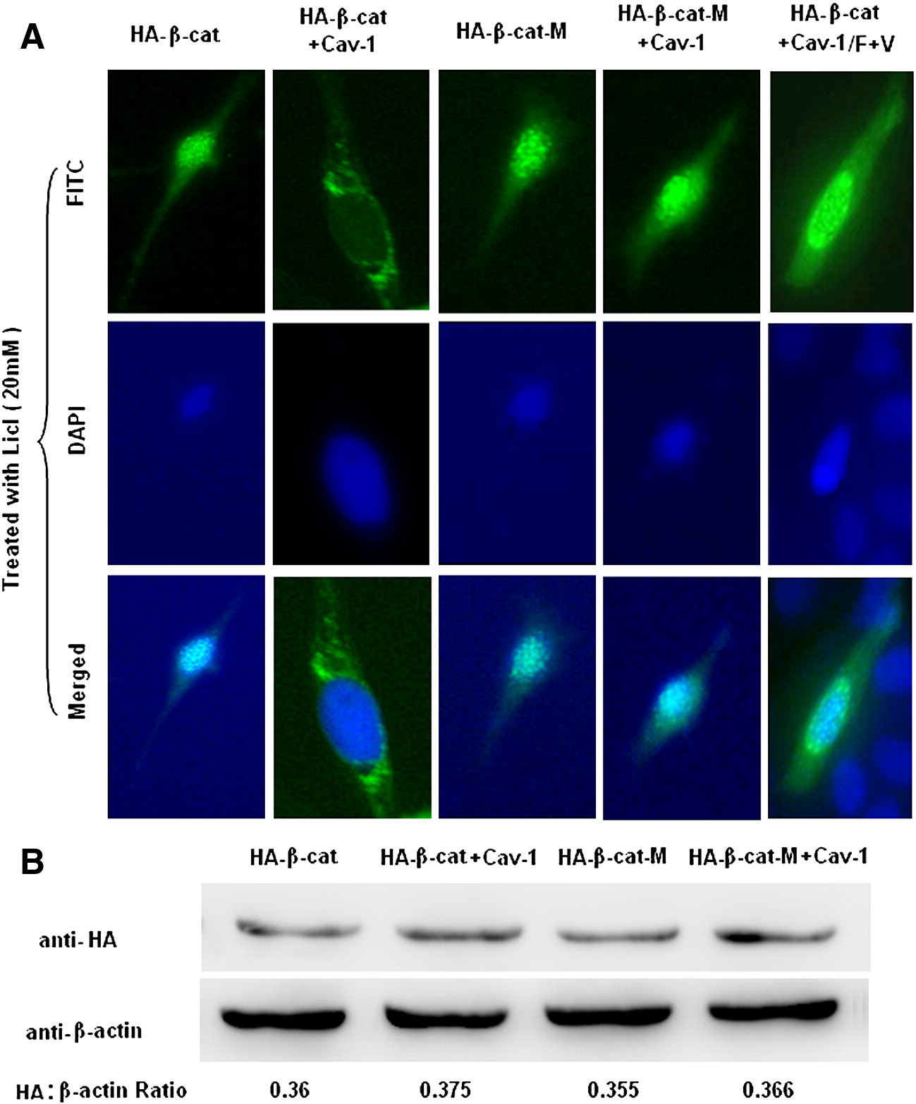
Fig. 6 Overexpression of Cav-1 blocks the nuclear translocation of active β-catenin. (A) ZF4 cells on 35 mm dishes were transfected with 600 ng of each plasmid expressing HA-β-cat plus pRC/CMV2, HA-β-cat plus Cav-1, HA-β-cat-M plus pRC/CMV2, HA-β-cat-M plus Cav-1, or HA-β-cat plus Cav-1/F + T. Transfected cells were then treated with 20 mM LiCl for 24 h. Cellular distribution of HA-β-cat or HA-β-cat-M was shown by location of FITC signal. Nuclei were stained with DAPI. (B) Effects of Cav-1 on degradation of β-catenin were detected by Western immunoblotting. ZF4 cells on 35 mm dishes were transfected with 600 ng of each plasmid expressing HA-β-cat plus pRC/CMV2, HA-β-cat plus Cav-1, HA-β-cat-M plus pRC/CMV2, or HA-β-cat-M plus Cav-1. Expression of β-actin was used as loading control.
Reprinted from Developmental Biology, 344(1), Mo, S., Wang, L., Li, Q., Li, J., Li, Y., Thannickal, V.J., and Cui. Z., Caveolin-1 regulates dorsoventral patterning through direct interaction with beta-catenin in zebrafish, 210-223, Copyright (2010) with permission from Elsevier. Full text @ Dev. Biol.