
IMAGE
Fig. S1
- ID
- ZDB-IMAGE-100706-33
- Publication
- Pujol-Martí et al., 2010 - Progressive neurogenesis defines lateralis somatotopy
- All Figures
- Figures for Pujol-Martí et al., 2010
Image
|
Figure Caption
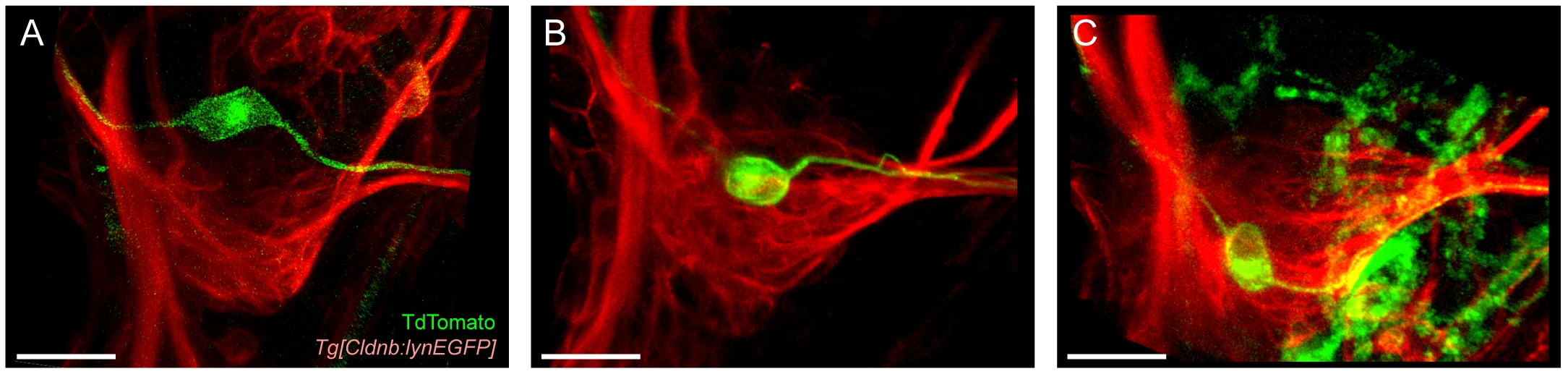
Fig. S1 A-C: Somata location in the posterior lateralis ganglion at 3.5 dpf of mem-TdTomato labeled neurons (green) with growth cones in the middle region of primI (A), trailing region of primI (B) or, behind primI (C). The red fluorescent signal is from Tg[CldnB:lynEGFP] transgenic line. All images are snapshots of the lateral view from three-dimensional reconstructions. Dorsal is towards top, anterior is towards left. Scale bars = 20 μm.
Acknowledgments
This image is the copyrighted work of the attributed author or publisher, and
ZFIN has permission only to display this image to its users.
Additional permissions should be obtained from the applicable author or publisher of the image.
Full text @ Dev. Dyn.