
|
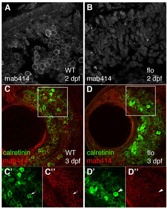
Fig. S1 Retinal cells in flo embryos contain disrupted nuclear pores, but still express a marker of neuronal differentiation and function. (A-D″) Nuclear pores and calretinin-positive cells were visualised in wild-type (A,C) and flo (B,D) retinae from 3 dpf by staining with mab414 (grey in A,B, red in C,D) and calretinin (green) sera. C′-D″ are enlargements of the single channels from the boxed regions in C and D, respectively. Calretinin-positive cells (green) from approximately the same retinal position have nuclei ringed uniformly by red mab414 staining (WT, C′,C″, arrow) or have aberrant nuclear pores (flo, D′,D″, arrowhead).