
|
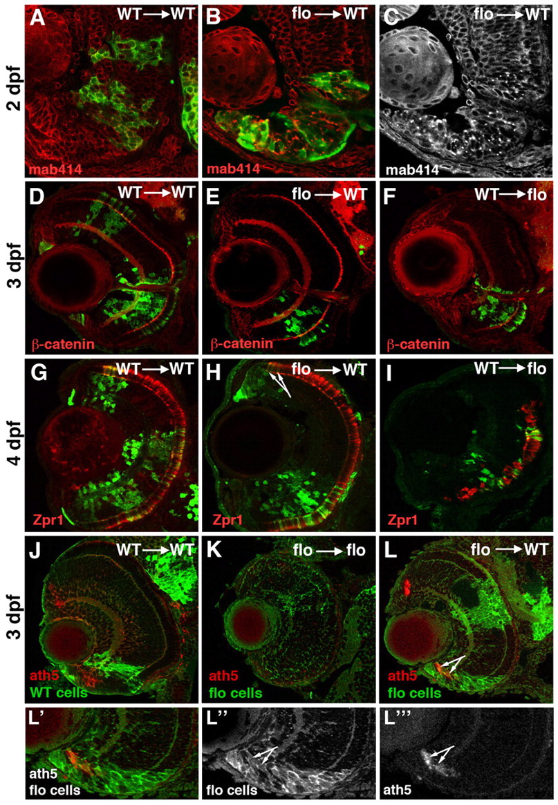
Fig. 6 flo cells survive and differentiate in a wild-type environment. (A-L″′) Frontal sections of zebrafish eyes at various ages labelled with antibodies as indicated bottom left; mab414 recognises the Nup107-160 subcomplex of nuclear pores, β-catenin highlights cell membranes and plexiform layers; zpr1 recognises cone photoreceptors. GFP-labelled cells from either wild-type (A,D,F,G,I,J) or flo (B,C,E,H,K,L) donor embryos were transplanted into wild-type (A-E,G,H,J,L) or flo (F,I,K) hosts. The arrows in H and L point to flo mutant photoreceptors and ath5-expressing cells emerging from the CMZ in wild-type eyes, respectively. See text for the specific numbers of mosaic eyes examined for each condition. ath5 expression (or lack thereof) in wild-type (J) and flo (K) retinae is shown for reference. (L′,L″,L″′) Single-channel magnifications from L illustrate flo CMZ cells in a wild-type environment.